

18F-PET/CT imaging of metastasis to the thyroid gland: Imaging findings and effect on patient management
- PMID: 30405862
- PMCID: PMC6217829
- DOI: 10.5430/jst.v7n2p7
18F-PET/CT imaging of metastasis to the thyroid gland: Imaging findings and effect on patient management
Abstract
Purpose: While metastasis to the thyroid from a primary cancer remote to the thyroid is uncommon, current imaging techniques have improved detection of these intrathyroid metastases. The purpose of this study was to evaluate the 18F-PET/CT appearance of intrathyroid metastases and assess the impact of detection on patient management.
Methods: The 18F-PET/CT appearance of intrathyroid metastasis, including standardized uptake value (SUV), disease extent, and the effect on patient management following diagnosis were retrospectively reviewed. Inclusion criteria included 18F-PET/CT imaging and diagnosis of the intrathyroid metastasis matching the remote primary tumor.
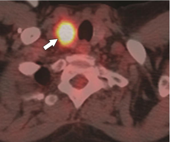
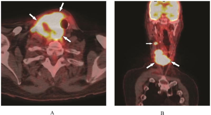
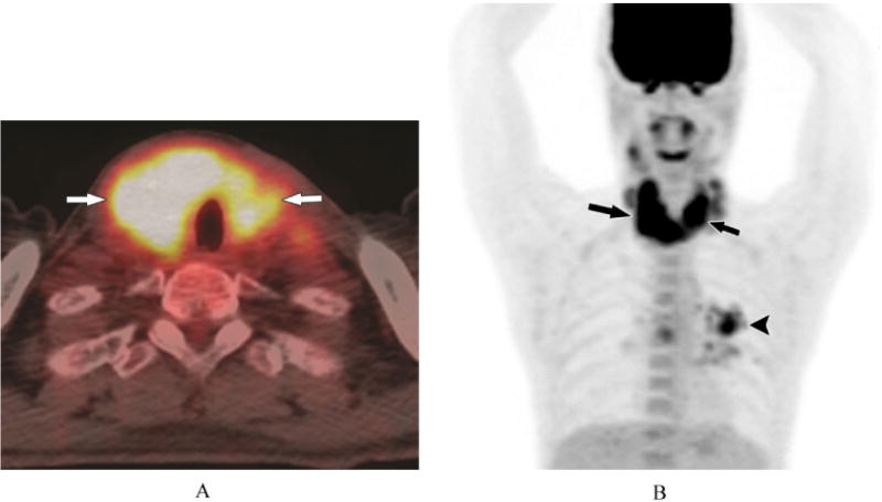
Results: Intrathyroid metastasis were detected in 24 patients. The intrathyroid metastases presented on 18F-PET/CT as focal nodular uptake (n = 21), multiple nodular uptake (n = 2), or diffuse uptake/infiltration of the thyroid gland (n = 1). The SUV ranged between 3.9 and 42 (median 12.5 ± 7.5); in 2 patients, the FDG-avidity was minimal. On 18F-PET/CT, distant metastases were present outside the neck (n = 18), or limited to the neck (n = 6). In 2 of these 6 patients, the thyroid was the only site of metastatic disease. Due to the metastatic disease, the therapy was changed in 23 of 24 patients; 1 patient was lost to follow-up.
Conclusion: In any patient with a previous or current history of an extrathyroid malignancy, an 18FDG-avid thyroid mass or diffuse infiltration of the thyroid on 18F-PET/CT should be considered a potential intrathyoid metastasis until proven otherwise. Knowledge of an intrathyroid metastasis may impact patient management, especially if the thyroid or neck are the only sites of metastatic disease.
Keywords: Metastasis; PET/CT; Standardized uptake value (SUV); Thyroid.
Conflict of interest statement
Conflicts of Interest Disclosure The authors declare that there is no conflict of interest statement.
Figures

Similar articles
-
Sonographic Evaluation of Intrathyroid Metastases.J Ultrasound Med. 2017 Jan;36(1):69-76. doi: 10.7863/ultra.16.02033. Epub 2016 Dec 7. J Ultrasound Med. 2017. PMID: 27925648
-
More advantages in detecting bone and soft tissue metastases from prostate cancer using 18F-PSMA PET/CT.Hell J Nucl Med. 2019 Jan-Apr;22(1):6-9. doi: 10.1967/s002449910952. Epub 2019 Mar 7. Hell J Nucl Med. 2019. PMID: 30843003
-
Focal thyroid lesions incidentally identified by integrated 18F-FDG PET/CT: clinical significance and improved characterization.J Nucl Med. 2006 Apr;47(4):609-15. J Nucl Med. 2006. PMID: 16595494
-
Prevalence and significance of thyroid uptake detected by ¹⁸F-FDG PET.Endocrine. 2011 Oct;40(2):297-302. doi: 10.1007/s12020-011-9470-5. Epub 2011 Apr 20. Endocrine. 2011. PMID: 21505891
-
Detection of thyroiditis on PET/CT imaging: a systematic review.Hormones (Athens). 2020 Sep;19(3):341-349. doi: 10.1007/s42000-020-00178-x. Epub 2020 Feb 10. Hormones (Athens). 2020. PMID: 32037486
Cited by
-
Rare Metastasis of Gastric Cancer to the Thyroid Gland: A Case Report and Review of Literature.Front Surg. 2021 Oct 8;8:731673. doi: 10.3389/fsurg.2021.731673. eCollection 2021. Front Surg. 2021. PMID: 34692762 Free PMC article.
-
Case report: Report of a rare encounter: metastasis of renal cell carcinoma to the thyroid.Front Oncol. 2024 Apr 23;14:1350043. doi: 10.3389/fonc.2024.1350043. eCollection 2024. Front Oncol. 2024. PMID: 38715782 Free PMC article.
-
Case report: Thyroid metastasis from hepatocellular carcinoma: a rare case with diffuse solid occupancy and unusual imaging findings.Front Oncol. 2024 Jul 10;14:1360734. doi: 10.3389/fonc.2024.1360734. eCollection 2024. Front Oncol. 2024. PMID: 39050581 Free PMC article.
-
Metastases to the Thyroid Gland: What Can We Do?Cancers (Basel). 2022 Jun 19;14(12):3017. doi: 10.3390/cancers14123017. Cancers (Basel). 2022. PMID: 35740683 Free PMC article. Review.
-
Role of Imaging in Clinically Occult Isolated Intrathyroidal Metastasis from Squamous Cell Carcinoma of Tongue: An Unusual Case Series.Indian J Nucl Med. 2018 Oct-Dec;33(4):326-330. doi: 10.4103/ijnm.IJNM_106_18. Indian J Nucl Med. 2018. PMID: 30386055 Free PMC article.
References
-
- Berge T, Lundberg S. Cancer in Malmo 1958-1969; An autopsy study. Acta Pathol Microbiol Scand Suppl. 1977;260:1–235. - PubMed
-
- Thorpe JD. Metastatic cancer in the thyroid gland: report of four cases. Surg Obstet Gynecol. 1954 Nov;62(11):574–6. - PubMed
-
- Shimaoka K, Sokal JE, Pickren JW. Metastatic neoplasms in the thyroid gland: pathological and clinical findings. 1962 May-Jun;15:557–65. - PubMed
Grants and funding
LinkOut - more resources
Full Text Sources