. 2018 Oct 24;48(5):284-294.
doi: 10.5051/jpis.2018.48.5.284. eCollection 2018 Oct.

Estrogen reinforces barrier formation and protects against tumor necrosis factor alpha-induced barrier dysfunction in oral epithelial cells

Affiliations

Estrogen reinforces barrier formation and protects against tumor necrosis factor alpha-induced barrier dysfunction in oral epithelial cells

Yun Sik Choi et al. J Periodontal Implant Sci. .

Abstract

Purpose: Epithelial barrier dysfunction is involved in the pathophysiology of periodontitis and oral lichen planus. Estrogens have been shown to enhance the physical barrier function of intestinal and esophageal epithelia, and we aimed to investigate the effect of estradiol (E2) on the regulation of physical barrier and tight junction (TJ) proteins in human oral epithelial cell monolayers.

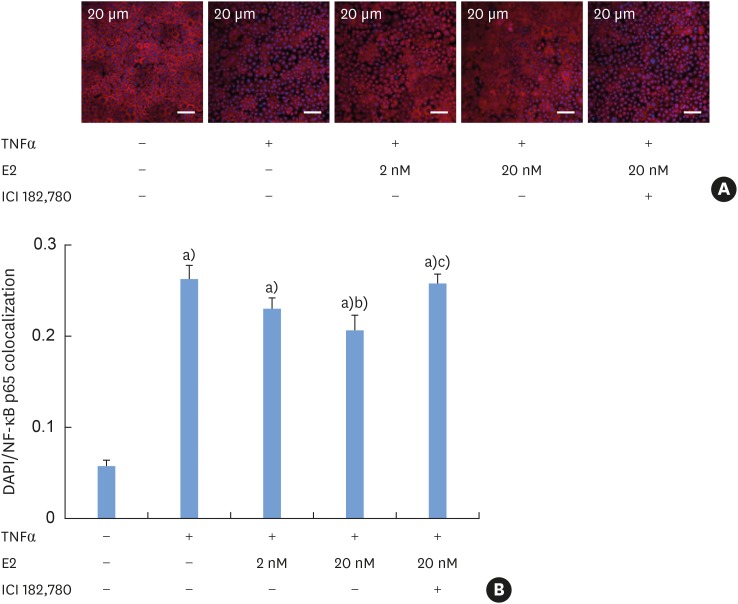
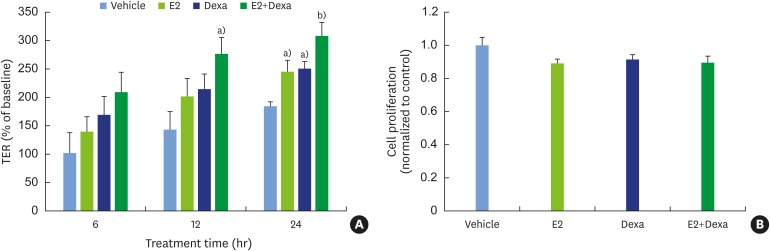
Methods: HOK-16B cell monolayers cultured on transwells were treated with E2, an estrogen receptor (ER) antagonist (ICI 182,780), tumor necrosis factor alpha (TNFα), or dexamethasone (Dexa), and the transepithelial electrical resistance (TER) was then measured. Cell proliferation was measured by the cell counting kit (CCK)-8 assay. The levels of TJ proteins and nuclear translocation of nuclear factor (NF)-κB were examined by confocal microscopy.

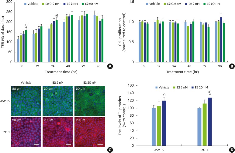
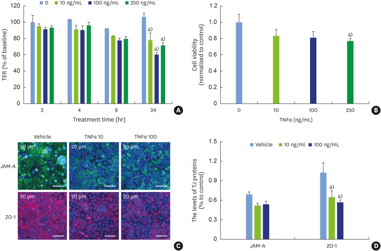
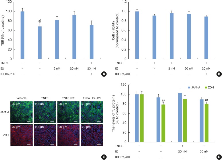
Results: E2 treatment increased the TER and the levels of junctional adhesion molecule (JAM)-A and zonula occludens (ZO)-1 in a dose-dependent manner, without affecting cell proliferation during barrier formation. Treatment of the tight-junctioned cell monolayers with TNFα induced decreases in the TER and the levels of ZO-1 and nuclear translocation of NF-κB. These TNFα-induced changes were inhibited by E2, and this effect was completely reversed by co-treatment with ICI 182,780. Furthermore, E2 and Dexa presented an additive effect on the epithelial barrier function.

Conclusions: E2 reinforces the physical barrier of oral epithelial cells through the nuclear ER-dependent upregulation of TJ proteins. The protective effect of E2 on the TNFα-induced impairment of the epithelial barrier and its additive effect with Dexa suggest its potential use to treat oral inflammatory diseases involving epithelial barrier dysfunction.

Keywords: Epithelial cells; Junctional adhesion molecule A; NF-kappa B; Tight junctions.


Conflict of interest statement

Conflict of Interest: No potential conflict of interest relevant to this article was reported.

Figures

Figure 1
Figure 1. Facilitation of barrier formation by E2 in oral epithelial cell monolayers. (A) Immortalized human oral keratinocyte (HOK-16B) cells seeded on the membrane of a transwell 2-chamber tissue culture system were treated with various concentrations of E2 during barrier formation. The TER was measured at the indicated time points and expressed as percentage of the baseline (0 hours). (B) HOK-16B cells plated onto 96-well plates in parallel were treated and cultured in the same way. The proliferation of HOK-16B cells was measured using the CCK-8 assay kit at the indicated time points. The measured absorbances are expressed as the relative index compared to the vehicle control at each time point. (C) HOK-16B cells plated on collagen coated cover slips were treated with 2 or 20 nM E2 for 24 hours. After fixation, cells were stained for JAM-A and ZO-1 and examined by confocal microscopy with serial z-sections. Images with a maximal intensity that combined the serial z-sections are shown. (D) The fluorescence intensities of JAM-A and ZO-1 were analyzed and normalized to the fluorescence intensity of Hochest 33342.
E2: estradiol, TER: transepithelial electrical resistance, CCK: cell counting kit, JAM: junctional adhesion molecule, ZO: zonula occludens, TJ: tight junction. a)P<0.05 versus vehicle control (2-tailed non-paired Student's t-test).
Figure 2
Figure 2. Disruption of the physical barrier by TNFα. Tight-junctioned monolayers of human oral keratinocyte (HOK-16B) cells were treated with various concentrations of TNFα for 24 hours. (A) The TER was measured at the indicated time points. (B) Cell viability was measured using the CCK-8 assay kit at 24 hours. (C) After fixation, cells were stained for JAM-A and ZO-1 and examined by confocal microscopy. (D) The fluorescence intensities of JAM-A and ZO-1 were analyzed.
TNFα: tumor necrosis factor alpha, TER: transepithelial electrical resistance, CCK: cell counting kit, JAM: junctional adhesion molecule, ZO: zonula occludens, TJ: tight junction. a)P<0.05 versus 0 ng/mL (2-tailed non-paired Student's t-test).
Figure 3
Figure 3. Protective effect of E2 on the TNFα-induced damage in oral epithelial physical barrier. Tight-junctioned monolayers of human oral keratinocyte (HOK-16B) cells were pre-treated with either ICI 182,780 for 6 hours and/or E2 for 4 hours and then treated with 100 ng/mL TNFα for 24 hours. (A) The TER was measured at the end point and expressed as the percentage of baseline. (B) Cell viability was measured using the CCK-8 assay kit at 24 hours. (C) After fixation, cells were stained for JAM-A and ZO-1 and examined by confocal microscopy. (D) The fluorescence intensities of JAM-A and ZO-1 were analyzed.
E2: estradiol, TER: transepithelial electrical resistance, TNFα: tumor necrosis factor alpha, CCK: cell counting kit, JAM: junctional adhesion molecule, ZO: zonula occludens, TJ: tight junction. a)P<0.05 versus no treatment (1-way analysis of variance with the Tukey post hoc test).
Figure 4
Figure 4. Inhibition of the TNFα-induced nuclear translocation of NF-κB by E2 (A) Tight-junctioned monolayers of human oral keratinocyte (HOK-16B) cells were pre-treated with either ICI 182,780 for 6 hours and/or E2 for 4 hours and then treated with 100 ng/mL TNFα for 30 minutes. After fixation, cells were stained for NF-κB p65 and examined by confocal microscopy. (B) Co-localized signals of nucleus and NF-κB p65 were measured.
E2: estradiol, TNFα: tumor necrosis factor alpha. a)P<0.0001 versus no treatment; b)P<0.05 versus treatment with TNFα alone; c)P<0.01 versus treatment with TNFα and 20 nM E2 (1-way analysis of variance with the Tukey post hoc test).
Figure 5
Figure 5. Additive effect of E2 and Dexa on the barrier formation in oral epithelial cell monolayers (A) Human oral keratinocyte (HOK-16B) cells seeded on the membrane of a transwell 2-chamber tissue culture system were treated with 20 nM E2, 250 nM Dexa, or E2+Dexa during barrier formation. The TER was measured at the indicated time points. (B) The proliferation of HOK-16B cells at 24 h was measured using the CCK-8 assay kit.
E2: estradiol, Dexa: dexamethasone, TER: transepithelial electrical resistance, CCK: cell counting kit. a)P<0.05 and b)P<0.0001 versus vehicle control (1-way analysis of variance with the Tukey post hoc test).

Similar articles

Cited by

References

    1. Ivanov AI, Parkos CA, Nusrat A. Cytoskeletal regulation of epithelial barrier function during inflammation. Am J Pathol. 2010;177:512–524. - PMC - PubMed
    1. Belibasakis GN, Kast JI, Thurnheer T, Akdis CA, Bostanci N. The expression of gingival epithelial junctions in response to subgingival biofilms. Virulence. 2015;6:704–709. - PMC - PubMed
    1. Brunner PM, Leung DY, Guttman-Yassky E. Immunologic, microbial, and epithelial interactions in atopic dermatitis. Ann Allergy Asthma Immunol. 2018;120:34–41. - PMC - PubMed
    1. Turner JR. Intestinal mucosal barrier function in health and disease. Nat Rev Immunol. 2009;9:799–809. - PubMed
    1. Khalmuratova R, Park JW, Shin HW. Immune cell responses and mucosal barrier disruptions in chronic rhinosinusitis. Immune Netw. 2017;17:60–67. - PMC - PubMed