. 2018 Oct 8:6:1900611.
doi: 10.1109/JTEHM.2018.2869141. eCollection 2018.

An Adaptive Seismocardiography (SCG)-ECG Multimodal Framework for Cardiac Gating Using Artificial Neural Networks

Affiliations

An Adaptive Seismocardiography (SCG)-ECG Multimodal Framework for Cardiac Gating Using Artificial Neural Networks

Jingting Yao et al. IEEE J Transl Eng Health Med. .

Abstract

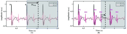
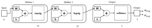
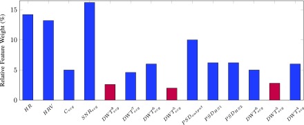
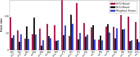
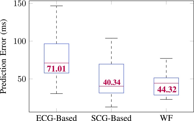
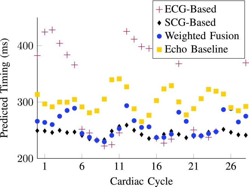
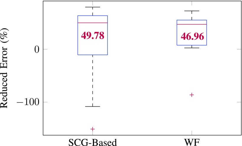
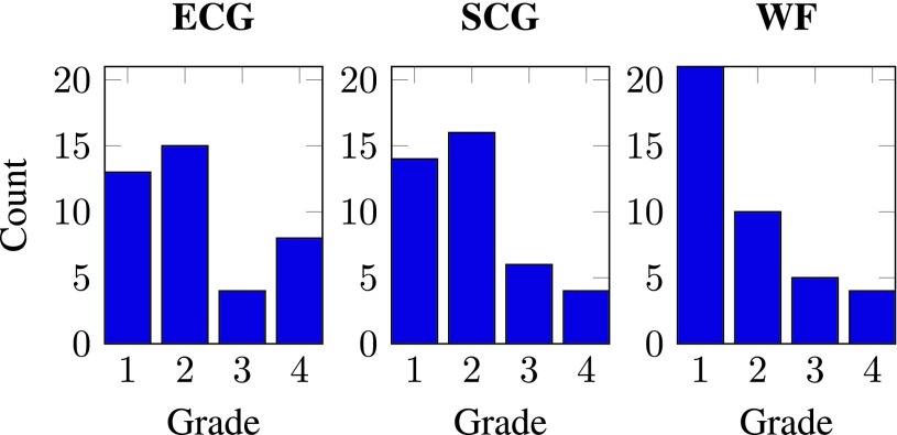
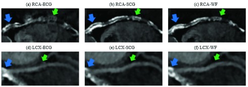
To more accurately trigger data acquisition and reduce radiation exposure of coronary computed tomography angiography (CCTA), a multimodal framework utilizing both electrocardiography (ECG) and seismocardiography (SCG) for CCTA prospective gating is presented. Relying upon a three-layer artificial neural network that adaptively fuses individual ECG- and SCG-based quiescence predictions on a beat-by-beat basis, this framework yields a personalized quiescence prediction for each cardiac cycle. This framework was tested on seven healthy subjects (age: 22-48; m/f: 4/3) and eleven cardiac patients (age: 31-78; m/f: 6/5). Seventeen out of 18 benefited from the fusion-based prediction as compared to the ECG-only-based prediction, the traditional prospective gating method. Only one patient whose SCG was compromised by noise was more suitable for ECG-only-based prediction. On average, our fused ECG-SCG-based method improves cardiac quiescence prediction by 47% over ECG-only-based method; with both compared against the gold standard, B-mode echocardiography. Fusion-based prediction is also more resistant to heart rate variability than ECG-only- or SCG-only-based prediction. To assess the clinical value, the diagnostic quality of the CCTA reconstructed volumes from the quiescence derived from ECG-, SCG- and fusion-based predictions were graded by a board-certified radiologist using a Likert response format. Grading results indicated the fusion-based prediction improved diagnostic quality. ECG may be a sub-optimal modality for quiescence prediction and can be enhanced by the multimodal framework. The combination of ECG and SCG signals for quiescence prediction bears promise for a more personalized and reliable approach than ECG-only-based method to predict cardiac quiescence for prospective CCTA gating.

Keywords: Artificial neural networks; cardiac gating; cardiac quiescence; computed tomography angiography; coronary angiography; echocardiography; electrocardiography; multimodal gating; seismocardiography.


Figures

Fig. 1.
Fig. 1.
Quiescence prediction methods. (A) The traditionally ECG-based prediction method; (B) Developed SCG-based prediction method. HS1 and HS2 are heart sound associated waveforms in systole and diastole, respectively , . The vertical dotted line is the quiescence derived from echocardiography which is considered as the baseline for quiescence in this study. Areas covered in grey contain succeeding unknown signals. The predicted quiescence, measured as a time formula image, is in reference to a cardiac feature within the upcoming cardiac cycle. As a demonstration we review predicting quiescence in diastole. Predicting formula image from HS2 involves less uncertainty than that from formula image using R-peak of ECG, therefore SCG-based prediction can potentially predict quiescence more accurately.
Fig. 2.
Fig. 2.
Three-layer ANN configuration . The input is a set of features consisting of 11 single-valued entries linked with two hidden layers with threshold functions tansig and logsig, each consisting of 10 neurons. formula image are configuration parameters representing the weights and bias. Two softmax output neurons in the output layer generate 2 values corresponding to the predicted probabilities, referred to as weights, of ECG- and SCG-based gating in the weighted fusion, WF.
Fig. 3.
Fig. 3.
Relative feature weight (%) evaluated by the neighborhood component analysis. The three features in plum bars demonstrated less importance in distinguishing ECG and SCG signals and consequently were discarded.
Fig. 4.
Fig. 4.
Quiescence prediction error (milliseconds) of different cardiac gating modalities. The overall prediction error across all subjects associated with ECG-, SCG- and WF-based method are 76.15 ms, 48.30 ms and 43.95 ms, respectively. For each subject, the optimal gating modality, either ECG, SCG or WF, is selected based on the least error (ms). Except for subject P2, all subjects demonstrate less prediction error using the WF- or SCG-based prediction for cardiac gating.
Fig. 5.
Fig. 5.
Box plot of quiescence prediction error (milliseconds) of all 18 subjects. On each box, the central mark indicates the median (value in red), and the bottom and top edges of the box indicate the 25th and 75th percentiles, respectively. No outlier was observed. ECG-based prediction resulted in the most error. WF and SCG-based predictions are comparable. The smallest variability is seen in the prediction error associated with WF.
Fig. 6.
Fig. 6.
A subset of predicted temporal quiescence derived from different gating modalities for patient P11. Overall, WF gating is the optimal gating modality for P11 according to the average error presented in Fig. 4.
Fig. 7.
Fig. 7.
Box plot of percentage of error reduction (%) against quiescence prediction error from ECG-based prediction across the 18 subjects. On each box, the central mark indicates the median (value in red), and the bottom and top edges of the box indicate the 25th and 75th percentiles, respectively. The outliers are plotted individually using the ‘+’ symbol. WF and SCG-based predictions are comparable and can reduce more percent of prediction errors than cohort-specific echocardiography. But the variability in the reduced error associated with WF is smaller than SCG.
Fig. 8.
Fig. 8.
Histograms of the diagnostic quality grades. Four point Likert response scale: 1 = excellent, 2 = good, 3 = adequate, 4 = non-diagnostic.
Fig. 9.
Fig. 9.
Comparison of the diagnostic quality of CCTA images reconstructed at quiescent phases derived from different gating modalities. CCTA data presented are from patient P11. Blue arrows point to one example of calcification. Green arrows point to motion artifacts. Compared to ECG-phases, the SCG-selected phases in (b) and (e), and WF-selected phases in (c) and (f) demonstrate sharper outline of the RCA and LCX. Calcification in the RCA is also more sharply defined by SCG- and WF-selected phases. Significant motion artifacts rendered the pointed (green arrows) regions of the RCA and LCX non-diagnostic for ECG-selected quiescent phases.

Similar articles

Cited by

References

    1. World Health Organization. (2017). Cardiovascular Disease. [Online]. Available: http://www.who.int/mediacentre/factsheets/fs317/en/
    1. Ricciardi M. J., Beohar N., and Davidson C. J., “Cardiac catheterization and coronary angiography,” in Essential Cardiology. Totowa, NJ, USA: Human Press, 2005, pp. 197–219.
    1. Desjardins B. and Kazerooni E. A., “ECG-gated cardiac CT,” Amer. J. Roentgenol., vol. 182, no. 4, pp. 993–1010, Apr. 2004. - PubMed
    1. Ladapo J. A.et al., “Clinical outcomes and cost-effectiveness of coronary computed tomography angiography in the evaluation of patients with chest pain,” J. Amer. College Cardiol., vol. 54, no. 25, pp. 2409–2422, 2009. - PubMed
    1. Priest V. L., Scuffham P. A., Hachamovitch R., and Marwick T. H., “Cost-effectiveness of coronary computed tomography and cardiac stress imaging in the emergency department: A decision analytic model comparing diagnostic strategies for chest pain in patients at low risk of acute coronary syndromes,” Cardiovascular Imag., vol. 4, no. 5, pp. 549–556, 2011. - PubMed