. 2018 Oct 10:2018:1754085.
doi: 10.1155/2018/1754085. eCollection 2018.

Staurosporine Induces the Generation of Polyploid Giant Cancer Cells in Non-Small-Cell Lung Carcinoma A549 Cells

Affiliations

Staurosporine Induces the Generation of Polyploid Giant Cancer Cells in Non-Small-Cell Lung Carcinoma A549 Cells

Alexander Glassmann et al. Anal Cell Pathol (Amst). .

Abstract

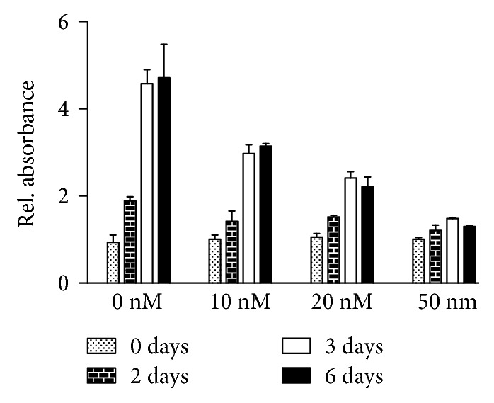
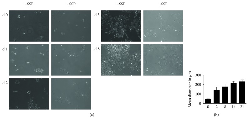
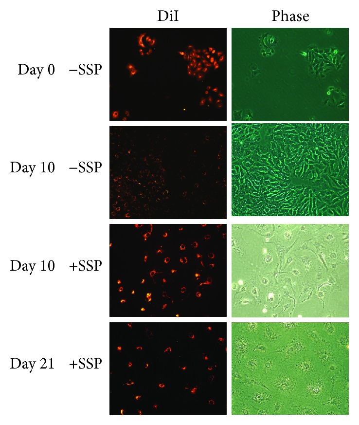
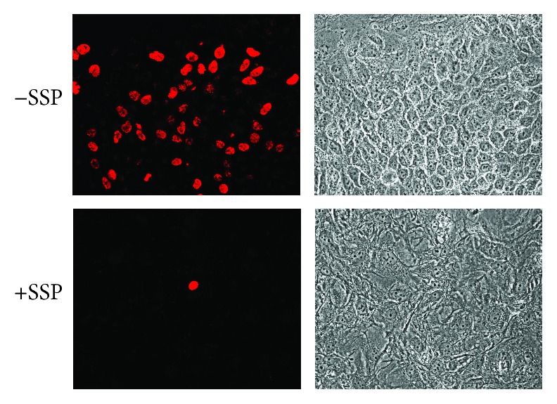
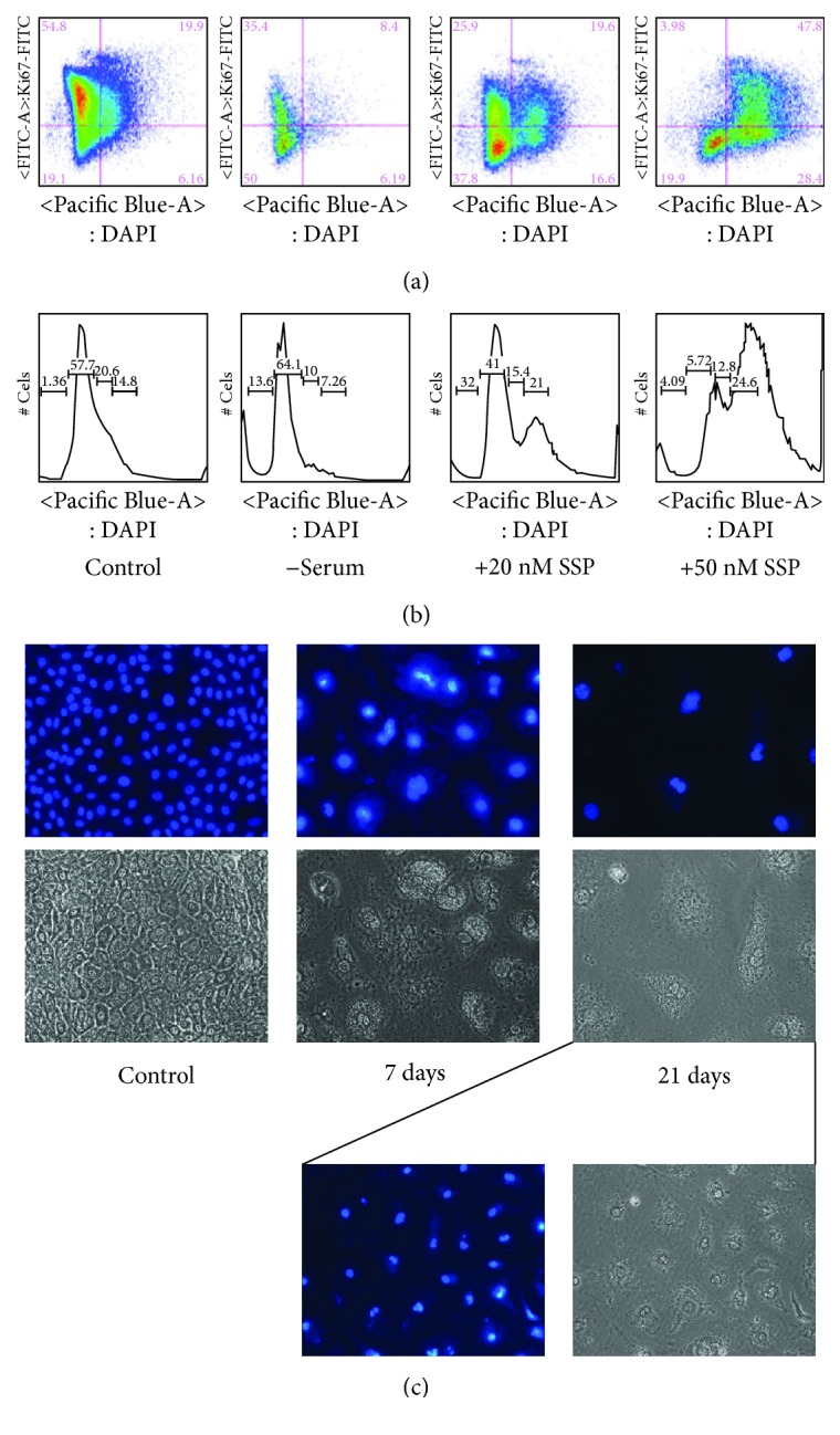
Cultivation of A549 non-small-cell lung carcinoma (NSCLC) cells in the presence of staurosporine (SSP) leads to a reduction or a lack of proliferation in a concentration-dependent manner. This inhibition of proliferation is accompanied by the generation of polyploid giant cancer cells (PGCCs) that are characterized by cell flattening, increased cell size, polyploidy, and polynucleation as determined by crystal violet staining, BrdU and DiI labelling, and flow cytometry as well as video time-lapse analysis. Continuous SSP treatment of A549 cells can preserve PGCCs for at least two months in a resting state. Upon removal of SSP, A549 PGCCs restart to divide and exhibit a proliferation pattern and cellular morphology indistinguishable from cells where PGCCs originally derived from. Thus, SSP-treated A549 cells represent a simple and reliable experimental model for the reversible generation of PGCCs and their subsequent experimental analysis.


Figures

Figure 1
Figure 1
Proliferation of A549 cells in the presence of SSP. A549 cells were cultivated with the indicated concentrations of SSP for 2, 3, or 6 days in 96-well plates. Cells were stained with crystal violet as specified in Materials and Methods. Absorbance of cells at day 0 was set at 1.0, and the other values were adjusted accordingly. Columns represent mean values + standard deviation (SD).
Figure 2
Figure 2
Micrographs (a) and size analysis (b) of A549 cells cultivated in the absence or presence of SSP. A549 cells were seeded at low density in Petri dishes and treated for up to 8 (a) or 21 (b) days without or with 50 nM SSP. In (b), at least 20 cells were analyzed for each time point. d: day(s).
Figure 3
Figure 3
Micrographs of DiI-labelled A549 cells cultivated in the absence or presence of SSP. A549 cells were labelled with DiI, seeded at low density in Petri dishes, and treated for up to 21 days without or with 50 nM SSP. Fluorescence and corresponding phase micrographs are shown.
Figure 4
Figure 4
SSP-dependent BrdU incorporation in A549 cells. Cells were seeded in 24-well plates at high density and cultivated for further 3 days in the absence or presence of 50 nM SSP. Cells were then pulsed for 4 h with BrdU. BrdU incorporation was visualized with anti-BrdU antibodies and secondary fluorescently labelled antibodies. Fluorescence and corresponding phase micrographs are shown.
Figure 5
Figure 5
SSP induces polyploidy and polynucleation in A549 cells. (a, b) FACS analysis of A549 cells that had been cultivated in normal growth medium with (control) or without (−serum) 10% FCS or in growth medium with 10% FCS containing 20 or 50 nM SSP. FACS plots are shown in (a), whereas in (b), the respective relative cell numbers in representative sections of micrographs in (a) are given on the y-axis. (c) DAPI staining to reveal the nuclear morphology of A549 cells that had been cultivated in the absence or presence of 50 nM SSP for 7 or 21 days. The corresponding phase micrographs are also given. In addition, a lower magnification of 21-day treated A549 cells is shown at the right bottom of (c) to reveal a larger cell number.
Figure 6
Figure 6
SSP-induced stop of proliferation is reversible. (a) Short- and long-time cultures of A549 cells in the absence or presence of 50 nM SSP. A549 cells were cultivated at the indicated time points with or without 50 nM SSP, stained with DAPI, and the cell number in selected areas was determined microscopically. Cell number at day zero (d 0) was set as 100%, and the other values were adjusted accordingly. (b) A549 cells that had been cultured in the presence of 50 nM SSP for three weeks were cultured in the absence of SSP for further three weeks. A colony of dividing cells in a dawn of still more flattened cells is indicated with an arrow. Fluorescence (DAPI label) and corresponding phase micrographs are shown.

Similar articles

Cited by

References

    1. Batlle E., Clevers H. Cancer stem cells revisited. Nature Medicine. 2017;23(10):1124–1134. doi: 10.1038/nm.4409. - DOI - PubMed
    1. Chen K., Huang Y. H., Chen J. L. Understanding and targeting cancer stem cells: therapeutic implications and challenges. Acta Pharmacologica Sinica. 2013;34(6):732–740. doi: 10.1038/aps.2013.27. - DOI - PMC - PubMed
    1. Wheatley D. Growing evidence of the repopulation of regressed tumours by the division of giant cells. Cell Biology International. 2008;32(9):1029–1030. doi: 10.1016/j.cellbi.2008.06.001. - DOI - PubMed
    1. Erenpreisa J., Cragg M. S. MOS, aneuploidy and the ploidy cycle of cancer cells. Oncogene. 2010;29(40):5447–5451. doi: 10.1038/onc.2010.310. - DOI - PubMed
    1. Weihua Z., Lin Q., Ramoth A. J., Fan D., Fidler I. J. Formation of solid tumors by a single multinucleated cancer cell. Cancer. 2011;117(17):4092–4099. doi: 10.1002/cncr.26021. - DOI - PMC - PubMed