

Setting Eyes on the Retinal Pigment Epithelium
- PMID: 30406103
- PMCID: PMC6207792
- DOI: 10.3389/fcell.2018.00145
Setting Eyes on the Retinal Pigment Epithelium
Abstract
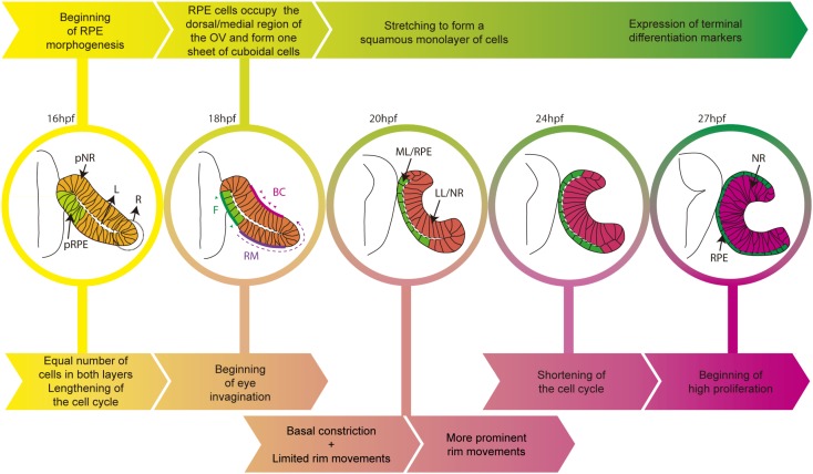
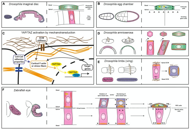
The neural component of the zebrafish eye derives from a small group of cells known as the eye/retinal field. These cells, positioned in the anterior neural plate, rearrange extensively and generate the optic vesicles (OVs). Each vesicle subsequently folds over itself to form the double-layered optic cup, from which the mature eye derives. During this transition, cells of the OV are progressively specified toward three different fates: the retinal pigment epithelium (RPE), the neural retina, and the optic stalk. Recent studies have shown that folding of the zebrafish OV into a cup is in part driven by basal constriction of the cells of the future neural retina. During folding, however, RPE cells undergo an even more dramatic shape conversion that seems to entail the acquisition of unique properties. How these changes occur and whether they contribute to optic cup formation is still poorly understood. Here we will review present knowledge on RPE morphogenesis and discuss potential mechanisms that may explain such transformation using examples taken from embryonic Drosophila tissues that undergo similar shape changes. We will also put forward a hypothesis for optic cup folding that considers an active contribution from the RPE.
Keywords: eye development; morphogenesis; optic cup; squamous epithelial cell; zebrafish (Danio rerio).
Figures
Similar articles
-
Stretching of the retinal pigment epithelium contributes to zebrafish optic cup morphogenesis.Elife. 2021 Sep 21;10:e63396. doi: 10.7554/eLife.63396. Elife. 2021. PMID: 34545806 Free PMC article.
-
Retinal pigment epithelium expansion around the neural retina occurs in two separate phases with distinct mechanisms.Dev Dyn. 2017 Aug;246(8):598-609. doi: 10.1002/dvdy.24525. Epub 2017 Jun 28. Dev Dyn. 2017. PMID: 28556369
-
Retinal Pigment Epithelium and Neural Retinal Progenitors Interact via Semaphorin 6D to Facilitate Optic Cup Morphogenesis.eNeuro. 2021 May 6;8(3):ENEURO.0053-21.2021. doi: 10.1523/ENEURO.0053-21.2021. Print 2021 May-Jun. eNeuro. 2021. PMID: 33811086 Free PMC article.
-
Eye morphogenesis and patterning of the optic vesicle.Curr Top Dev Biol. 2010;93:61-84. doi: 10.1016/B978-0-12-385044-7.00003-5. Curr Top Dev Biol. 2010. PMID: 20959163 Free PMC article. Review.
-
Cell fate decisions, transcription factors and signaling during early retinal development.Prog Retin Eye Res. 2022 Nov;91:101093. doi: 10.1016/j.preteyeres.2022.101093. Epub 2022 Jul 8. Prog Retin Eye Res. 2022. PMID: 35817658 Free PMC article. Review.
Cited by
-
Hippo effector, Yorkie, is a tumor suppressor in select Drosophila squamous epithelia.Proc Natl Acad Sci U S A. 2024 Sep 24;121(39):e2319666121. doi: 10.1073/pnas.2319666121. Epub 2024 Sep 17. Proc Natl Acad Sci U S A. 2024. PMID: 39288176 Free PMC article.
-
Ocular elongation and retraction in foveated reptiles.Dev Dyn. 2021 Nov;250(11):1584-1599. doi: 10.1002/dvdy.348. Epub 2021 May 1. Dev Dyn. 2021. PMID: 33866663 Free PMC article.
-
Cilia-associated wound repair mediated by IFT88 in retinal pigment epithelium.Sci Rep. 2023 May 21;13(1):8205. doi: 10.1038/s41598-023-35099-3. Sci Rep. 2023. PMID: 37211572 Free PMC article.
-
Identification of 4 novel human ocular coloboma genes ANK3, BMPR1B, PDGFRA, and CDH4 through evolutionary conserved vertebrate gene analysis.Genet Med. 2022 May;24(5):1073-1084. doi: 10.1016/j.gim.2021.12.014. Epub 2022 Jan 13. Genet Med. 2022. PMID: 35034853 Free PMC article.
-
Nf2 fine-tunes proliferation and tissue alignment during closure of the optic fissure in the embryonic mouse eye.Hum Mol Genet. 2020 Dec 18;29(20):3373-3387. doi: 10.1093/hmg/ddaa228. Hum Mol Genet. 2020. PMID: 33075808 Free PMC article.
References
Publication types
LinkOut - more resources
Full Text Sources
Molecular Biology Databases