

Initial Steps in Mammalian Autophagosome Biogenesis
- PMID: 30406104
- PMCID: PMC6206277
- DOI: 10.3389/fcell.2018.00146
Initial Steps in Mammalian Autophagosome Biogenesis
Abstract
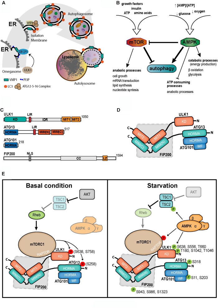
During the last decade, autophagy has been pointed out as a central process in cellular homeostasis with the consequent implication in most cellular settings and human diseases pathology. At present, there is significant data available about molecular mechanisms that regulate autophagy. Nevertheless, autophagy pathway itself and its importance in different cellular aspects are still not completely clear. In this article, we are focused in four main aspects: (a) Induction of Autophagy: Autophagy is an evolutionarily conserved mechanism induced by nutrient starvation or lack of growth factors. In higher eukaryotes, autophagy is a cell response to stress which starts as a consequence of organelle damage, such as oxidative species and other stress conditions. (b) Initiation of Autophagy; The two major actors in this signaling process are mTOR and AMPK. These multitasking protein complexes are capable to summarize the whole environmental, nutritional, and energetic status of the cell and promote the autophagy induction by means of the ULK1-Complex, that is the first member in the autophagy initiation. (c) ULK1-Complex: This is a highly regulated complex responsible for the initiation of autophagosome formation. We review the post-transductional modifications of this complex, considering the targets of ULK1. (d)The mechanisms involved in autophagosome formation. In this section we discuss the main events that lead to the initial structures in autophagy. The BECN1-Complex with PI3K activity and the proper recognition of PI3P are one of these. Also, the transmembrane proteins, such as VMP1 and ATG9, are critically involved. The membrane origin and the cellular localization of autophagosome biogenesis will be also considered. Hence, in this article we present an overview of the current knowledge of the molecular mechanisms involved in the initial steps of mammalian cell autophagosome biogenesis.
Keywords: AMPK; ULK1; VMP1; autophagy regulation; mTOR.
Figures
Similar articles
-
A Double Negative Feedback Loop between mTORC1 and AMPK Kinases Guarantees Precise Autophagy Induction upon Cellular Stress.Int J Mol Sci. 2019 Nov 7;20(22):5543. doi: 10.3390/ijms20225543. Int J Mol Sci. 2019. PMID: 31703252 Free PMC article.
-
The Emerging Roles of mTORC1 in Macromanaging Autophagy.Cancers (Basel). 2019 Sep 24;11(10):1422. doi: 10.3390/cancers11101422. Cancers (Basel). 2019. PMID: 31554253 Free PMC article. Review.
-
AMPK Inhibits ULK1-Dependent Autophagosome Formation and Lysosomal Acidification via Distinct Mechanisms.Mol Cell Biol. 2018 Apr 30;38(10):e00023-18. doi: 10.1128/MCB.00023-18. Print 2018 May 15. Mol Cell Biol. 2018. PMID: 29507183 Free PMC article.
-
ER-Targeted Beclin 1 Supports Autophagosome Biogenesis in the Absence of ULK1 and ULK2 Kinases.Cells. 2019 May 17;8(5):475. doi: 10.3390/cells8050475. Cells. 2019. PMID: 31108943 Free PMC article.
-
Autophagosome formation--the role of ULK1 and Beclin1-PI3KC3 complexes in setting the stage.Semin Cancer Biol. 2013 Oct;23(5):301-9. doi: 10.1016/j.semcancer.2013.05.007. Epub 2013 May 29. Semin Cancer Biol. 2013. PMID: 23727157 Review.
Cited by
-
Autophagy in health and disease: From molecular mechanisms to therapeutic target.MedComm (2020). 2022 Jul 10;3(3):e150. doi: 10.1002/mco2.150. eCollection 2022 Sep. MedComm (2020). 2022. PMID: 35845350 Free PMC article. Review.
-
SIRT1 and Autophagy: Implications in Endocrine Disorders.Front Endocrinol (Lausanne). 2022 Jul 14;13:930919. doi: 10.3389/fendo.2022.930919. eCollection 2022. Front Endocrinol (Lausanne). 2022. PMID: 35909524 Free PMC article. Review.
-
Targeting programmed cell death pathways: emerging therapeutic strategies for diabetic kidney disease.Front Endocrinol (Lausanne). 2025 Jun 11;16:1513895. doi: 10.3389/fendo.2025.1513895. eCollection 2025. Front Endocrinol (Lausanne). 2025. PMID: 40568564 Free PMC article. Review.
-
Autophagy-Mediated Exosomes as Immunomodulators of Natural Killer Cells in Pancreatic Cancer Microenvironment.Front Oncol. 2021 Feb 19;10:622956. doi: 10.3389/fonc.2020.622956. eCollection 2020. Front Oncol. 2021. PMID: 33680945 Free PMC article. Review.
-
Degradation of the Tumor Suppressor PDCD4 Is Impaired by the Suppression of p62/SQSTM1 and Autophagy.Cells. 2020 Jan 15;9(1):218. doi: 10.3390/cells9010218. Cells. 2020. PMID: 31952347 Free PMC article.
References
LinkOut - more resources
Full Text Sources
Miscellaneous