Case Reports
. 2018 Oct 22:5:260.
doi: 10.3389/fvets.2018.00260. eCollection 2018.

Presence of Probst Bundles Indicate White Matter Remodeling in a Dog With Corpus Callosum Hypoplasia and Dysplasia

Affiliations
Case Reports

Presence of Probst Bundles Indicate White Matter Remodeling in a Dog With Corpus Callosum Hypoplasia and Dysplasia

Adriano Wang-Leandro et al. Front Vet Sci. .

Abstract

Corpus callosum abnormalities (CCA) rarely occur in dogs and are related to hypo/adypsic hypernatremia and seizures. Hypoplasia and dysplasia of the corpus callosum (CC) with concomitant lobar holoprosencephaly is the most common variant. It is currently uncertain using conventional MRI if canine CCA reflects the failure of commissural fibers to develop or the failure of the commissural fibers to cross hemispheres. Diffusion tensor imaging was performed in a 4-year-old Staffordshire mix breed dog with CCA and an age-matched healthy Beagle. In comparison to the control dog, CC tractography of the affected dog depicted only axonal tracts corresponding to the temporal CC fibers. The cingulum bundles appeared supernumerary with unorganized architecture, extending into the ipsilateral cerebral cortex, and therefore strongly suggested homology to Probst bundles reported in humans with CCA. The presence of Probst bundles in canine CCA could represent compensatory neuroplasticity-mediated networking and may contribute the fair prognosis reported in affected dogs.

Keywords: DTI; adipsia; axonal bundles; canine; epilepsy; hypernatremia; seizures.


Figures
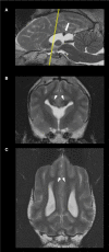
Figure 1
Figure 1
Sagittal (A), transverse, at the level of the optic chiasma (B), and dorsal, at the level of the cingulum (C), T2-weighted images of the 4 year-old, male Staffordshire Terrier mix with hypodipsia and seizures. The yellow line in (A) indicates the level at which the transverse plane is depicted. The corpus callosum (CC) is hypoplastic and dysplastic. The white arrow points to a discrete focal area of preserved thickness and white matter signal intensity within the splenium of the CC (A). The arrowheads point at the mildly enlarged cingula, bilaterally (C). The lateral ventricles and rostroventral segments of the frontal lobes are fused; there were no detectable fornix nor septum pellucidum (B).
Figure 2
Figure 2
Whole brain tractographies registered to T1-W images at the level of the caudal colliculi depicted in a dorsal plane and corpus callosum tractographies in the sagittal plane (rostral to the left) of a 4-year-old, male healthy Beagle (A,C) and the 4-year-old, male Staffordshire Terrier mix with corpus callosum abnormalities (CCA; B,D). The majority of axonal bundles crossing hemispheres are absent in the dog with CCA (white arrow; (B) in comparison to the healthy Beagle. Tractography of the CC revealed that only temporal fibers were present, corresponding to the discrete area of remaining white matter noticed in T2-weighted sequences (D) Color coding of tracts: Red, laterolateral axis; green ventrodorsal axis; blue, rostrocaudal axis. F Ant, frontal anterior; F Orb, frontal orbital; F Sup, frontal superior; Occ, occipital; Par, parietal; Temp, temporal.
Figure 3
Figure 3
Tractographies derived from regions of interest positioned at the cingulum of a 4-year-old, male healthy Beagle (A,C) and the 4-year-old, male Staffordshire Terrier mix with corpus callosum abnormalities (CCA; B,D). The tractographies are shown in the dorsal plane, registered to T1-W images at the level of the thalamus (A,B) and in the sagittal plane (rostral to the left; C,D). Supernumerary axonal bundles with unorganized morphology projecting to the ipsilateral cortex are present in the dog affected by CCA in comparison to the control dog. Color coding of tracts: Red, laterolateral axis; green, ventrodorsal axis; blue, rostrocaudal axis.

Similar articles

Cited by

References

    1. Goncalves R, Volk H, Smith PM, Penderis J, Garosi L, MacKillop E, et al. . Corpus callosal abnormalities in dogs. J Vet Internal Med. (2014) 28:1275–9. 10.1111/jvim.12371 - DOI - PMC - PubMed
    1. Sullivan SA, Harmon BG, Purinton PT, Greene CE, Glerum LE. Lobar holoprosencephaly in a Miniature Schnauzer with hypodipsic hypernatremia. J Am Vet Med Assoc. (2003) 223:1783–7. 10.2460/javma.2003.223.1783 - DOI - PubMed
    1. Benezit A, Hertz-Pannier L, Dehaene-Lambertz G, Monzalvo K, Germanaud D, Duclap D, et al. . Organising white matter in a brain without corpus callosum fibres. Cortex (2015) 63:155–71. 10.1016/j.cortex.2014.08.022 - DOI - PubMed
    1. Filippi CG, Cauley KA. Lesions of the corpus callosum and other commissural fibers: diffusion tensor studies. Semin Ultrasound CT MR (2014) 35:445–58. 10.1053/j.sult.2014.06.004 - DOI - PubMed
    1. Poretti A, Meoded A, Rossi A, Raybaud C, Huisman TA. Diffusion tensor imaging and fiber tractography in brain malformations. Pediatric Radiol. (2013) 43:28–54. 10.1007/s00247-012-2428-9 - DOI - PubMed

Publication types

LinkOut - more resources