. 2018 Oct 23;5(5):ENEURO.0367-18.2018.
doi: 10.1523/ENEURO.0367-18.2018. eCollection 2018 Sep-Oct.

Zic4-Lineage Cells Increase Their Contribution to Visual Thalamic Nuclei during Murine Embryogenesis If They Are Homozygous or Heterozygous for Loss of Pax6 Function

Affiliations

Zic4-Lineage Cells Increase Their Contribution to Visual Thalamic Nuclei during Murine Embryogenesis If They Are Homozygous or Heterozygous for Loss of Pax6 Function

Ziwen Li et al. eNeuro. .

Abstract

Our aim was to study the mechanisms that contribute to the development of discrete thalamic nuclei during mouse embryogenesis (both sexes included). We characterized the expression of the transcription factor coding gene Zic4 and the distribution of cells that expressed Zic4 in their lineage. We used genetic fate mapping to show that Zic4-lineage cells mainly contribute to a subset of thalamic nuclei, in particular the lateral geniculate nuclei (LGNs), which are crucial components of the visual pathway. We observed that almost all Zic4-lineage diencephalic progenitors express the transcription factor Pax6 at variable location-dependent levels. We used conditional mutagenesis to delete either one or both copies of Pax6 from Zic4-lineage cells. We found that Zic4-lineage cells carrying either homozygous or heterozygous loss of Pax6 contributed in abnormally high numbers to one or both of the main lateral geniculate nuclei (LGNs). This could not be attributed to a change in cell production and was likely due to altered sorting of thalamic cells. Our results indicate that positional information encoded by the levels of Pax6 in diencephalic progenitors is an important determinant of the eventual locations of their daughter cells.

Keywords: Pax6; Zic4; lateral geniculate nucleus; prethalamus; thalamus.


Figures
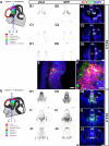
Figure 1.
Figure 1.
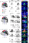
Expression of Zic4 and activity of Zic4Cre in Zic4Cre+/−;RCE+/− mouse brain coronal sections at E11.5 and E12.5. A, G, Schematics of E11.5 and E12.5 mouse brain demarcating major subdivisions and sectioning planes. B1–F, H1–J3, Zic4 fluorescent in situ hybridization (monochrome in B1, C1, D1, H1, I1, J1 or red in B3, C3, D3, E, F, H3, I3, J3) and immunohistochemistry for GFP reporter of Zic4Cre activity (monochrome in B2, C2, D2, H2, I2, J2 or green in B3, C3, D3, E, F, H3, I3, J3) with DAPI counterstaining (blue). Box in D3 is shown in E; box in E is shown in F. Scale bars: 250 µm (B1–D3, H1–J3), 125 µm (E), 25 µm (F). Cx, cortex; cp, choroid plexus; MGE, medial ganglionic eminence; LGE, lateral ganglionic eminence; th, thalamus; pth, prethalamus; emt, eminentia thalami; epi, epithalamus.
Figure 2.
Figure 2.
Expression of Zic4 and activity of Zic4Cre in Zic4Cre+/−;RCE+/− mouse brain coronal sections at E13.5, E14.5, and E15.5. A, E, I, Schematics of E13.5, E14.5, and E15.5 mouse brain demarcating major subdivisions and sectioning planes. B1–D3, F1–H3, J1–L3, Zic4 fluorescent in situ hybridization (monochrome in B1, C1, D1, F1, G1, H1, J1, K1, L1 or red in B3, C3, D3, F3, G3, H3, J3, K3, L3) and immunohistochemistry for GFP reporter of Zic4Cre activity (monochrome in B2, C2, D2, F2, G2, H2, J2, K2, L2 or green in B3, C3, D3, F3, G3, H3, J3, K3, L3) with DAPI counterstaining (blue). Scale bars: 250 µm. vLGN, ventral lateral geniculate nucleus; th, thalamus; pth, prethalamus.
Figure 3.
Figure 3.
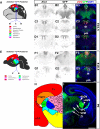
Expression of Zic4 and activity of Zic4Cre in Zic4Cre+/−;RCE+/− mouse brain coronal sections at E16.5, E18.5, and P0. A, E, H, Schematics of E16.5, E18.5, and P0 mouse brain demarcating major subdivisions and nuclei and sectioning planes. B1–D3, F1–G3, Zic4 fluorescent in situ hybridization (monochrome in B1, C1, D1, F1, G1 or red in B3, C3, D3, F3, G3) and immunohistochemistry for GFP reporter of Zic4Cre activity (monochrome in B2, C2, D2, F2, G2 or green in B3, C3, D3, F3, G3, I) with DAPI counterstaining (blue). Scale bars: 250 µm (B1–G3), 500 µm (I). Cx, cortex; dLGN, dorsal lateral geniculate nucleus; Hippo, hippocampus; LD, lateral dorsal nucleus; LH, lateral habenula; LP, lateral posterior nucleus; MD, medial dorsal nucleus; MH, medial habenula; PO, posterior nucleus; pth, prethalamus; PV, paraventricular nucleus; RE, nucleus reunions; RH, rhomboid nucleus; SPF, subparafascicular thalamic nucleus; th, thalamus; VAL, ventral anterolateral nucleus; vLGN, ventral lateral geniculate nucleus; VM, ventromedial nucleus; VPL, ventral posterolateral nucleus; VPM = ventral posteromedial nucleus; vTel, ventral telencephalon; ZI, zona incerta. H, Adapted from Allen Brain Atlas (http://developingmouse.brain-map.org/).
Figure 4.
Figure 4.
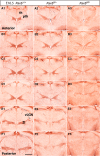
Pax6 expression in control and Pax6 mutant embryos at E12.5. Immunohistochemistry for Pax6 on a set of sections cut through the diencephalon in similar planes of section to those shown in Figure 1. Boxed areas in B1–B3 are enlarged in F1–F3. ZLI, zona limitans intrathalamica; epi, epithalamus; th, thalamus; pth, prethalamus; emt, eminentia thalami; h, hypothalamus. Scale bars: 500 µm (A1–E3), 50 µm (F1–F3).
Figure 5.
Figure 5.
Pax6 expression in control and Pax6 mutant embryos at E13.5. Immunohistochemistry for Pax6 on a set of sections cut through the diencephalon in similar planes of section to those shown in Figure 2. Boxed areas in A1–B3 are enlarged in F1–G3. vLGN, ventral lateral geniculate nucleus; epi, epithalamus; th, thalamus; pth, prethalamus; emt, eminentia thalami; h, hypothalamus. Scale bars: 500 µm (A1–E3), 50 µm (F1–F3).
Figure 6.
Figure 6.
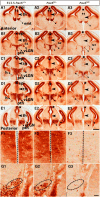
Pax6 expression in control and Pax6 mutant embryos at E14.5. (A1–G3) Immunohistochemistry for Pax6 on a set of sections cut through the diencephalon in similar planes of section to those shown in Figure 2, arranged from anterior (A1–A3) to posterior (G1–G3). ZI, zona incerta; vLGN, ventral lateral geniculate nucleus; th, thalamus; pth, prethalamus; emt, eminentia thalami. Scale bar: 500 µm.
Figure 7.
Figure 7.
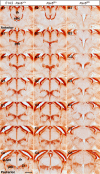
Pax6 expression in control and Pax6 mutant embryos at E16.5. (A1–F3)Immunohistochemistry for Pax6 on a set of sections cut through the diencephalon in similar planes of section to those shown in Figure 3, arranged from anterior (A1–A3) to posterior (F1–F3). ZI, zona incerta; vLGN, ventral lateral geniculate nucleus; th, thalamus; pth, prethalamus. Scale bar: 500 µm.
Figure 8.
Figure 8.
Diencephalic Zic4-lineage cells express Pax6. Double-immunohistochemistry for GFP (Zic4-lineage cells) and Pax6 at E11.5. Area outlined in B is enlarged in C. Asterisks in C: yellow, examples of double-labeled cells; green, examples of cells labeled only with GFP; red, examples of cells labeled only for Pax6. Scale bars: 250 µm (A), 50 µm (B), 10 µm (C).
Figure 9.
Figure 9.
Birthdates of cells in the vLGN, dLGN, and VP. A–D, Immunohistochemistry at P0 with markers designed to help delineate borders of the thalamic nuclei analyzed here. Scale bar: 100 µm. E, F, Example of BrdU and GFP immunohistochemistry in vLGN. Scale bars: 100 µm (E), 10 µm (F). G, Proportions of all cells in each nucleus that were BrdU+ after injection with BrdU at E10.5–E13.5. H, Proportions of GFP-expressing cells in each nucleus that were BrdU+ after injection with BrdU at E10.5–E13.5. Data points are from individual animals. Mean ± SEM are shown in each case. One-way ANOVA returned significant effects of age in all cases: (G) vLGN F(3,12) = 7.729, p = 0.0039; dLGN F(3,12) = 13.56, p = 0.0004; VP F(3,12) = 20.76, p < 0.0001; (H) vLGN F(3,12) = 8.282, p = 0.0030; dLGN F(3,12) = 15.41, p = 0.0002; VP F(3,12) = 23.26, p < 0.0001. Holm–Sidak’s multiple comparisons tests were performed following one-way ANOVA: *p < 0.05; **p < 0.01; ***p < 0.001.
Figure 10.
Figure 10.
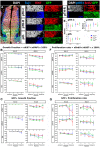
Effects of Pax6 mutation in Zic4-lineage cells on early diencephalic progenitor proliferation. A, Triple immunostaining for Tuj1, Ki67, and GFP. Three regions of interest were selected for analysis, one midway through pTH-C, one in pTH-R and one in prethalamus (pth). Scale bars: 50 µm. B, Average growth fractions (±SEM) in five equally spaced sections through pTH-C and pTH-R and four through prethalamus at E11.5 and E12.5; color coding for genotypes as in E. Two-way ANOVA detected significant effects of genotype only in pTH-C (F(2,30) = 9.109, p = 0.0008) and pTH-R (F(2,30) = 11.97, p = 0.0002) at E12.5 (n = 3 embryos of each genotype), with Pax6fl/fl embryos showing lower growth fractions at all levels (post hoc Tukey’s multiple comparisons test, p < 0.01 at all positions). C, Average growth fraction for GFP+ cells only. There were no significant effects of genotype. D, An example of triple immunostaining for pHH3, BrdU, and GFP in one region of interest. E, Mean (±SEM) counts of the total numbers of pHH3+ cells in all sampling regions from each domain for each genotype. There were no significant effects of genotype. F, Average proliferation rates (±SEM) in five equally spaced sections through pTH-C and pTH-R and four through prethalamus at E11.5 and E12.5; color coding for genotypes as in E. Two-way ANOVA showed a significant effect of genotype in pTH-R at E11.5 (F(2,30) = 4.206, p = 0.0245), with lower values in Pax6fl/+ embryos than in the other two genotypes. G, Average proliferation rates for GFP+ cells only. There were no significant effects of genotype.
Figure 11.
Figure 11.
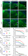
Pax6 mutation in Zic4-lineage cells increases their contribution to vLGN and dLGN. A–C, Sections from rostral to caudal through the diencephalon of P0 Zic4Cre;Pax6+/+, Zic4Cre;Pax6fl/+, and Zic4Cre;Pax6fl/fl embryos. Scale bar: 750 µm. D–F, Total numbers of cells in vLGN, dLGN, and VP in four P0 pups of each genotype; individual values, mean ± SEM are shown. There were no significant effects of genotype. G–I, Proportions of GFP+ (i.e., Zic4-lineage) cells in sections equally spaced through the vLGN, dLGN, and VP of P0 Zic4Cre;Pax6+/+, Zic4Cre;Pax6fl/+, and Zic4Cre;Pax6fl/fl pups. In vLGN and dLGN, two-way ANOVA showed significant effects of genotype: vLGN, F(2,90) = 23.98, p < 0.0001; dLGN, F(2,60) = 140.4, p < 0.0001. In VP, there were no significant effects. Data are plotted using mean and SEM using at least three pups of each genotype (n = 4 for vLGN and n = 3 for dLGN and VP).
Figure 12.
Figure 12.
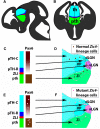
Model. A, B, Sections through the embryonic thalamus (th) and prethalamus (pth) showing the locations of the ZLI (red) and pTH-R (dark blue). C, D, Normally, Zic4-lineage cells from progenitors in the ZLI and pTH-R, which express little or no Pax6, contribute to the vLGN. Zic4-lineage cells from progenitors in rostral pTH-C (i.e., close to pTH-R), whose levels of Pax6 are relatively low, contribute to dLGN. Zic4-lineage cells in prethalamus (pth), which express Pax6 at high levels in both progenitors and postmitotic neurons, contribute to prethalamic regions including the ZI. E, F, Mutant Zic4-lineage cells concentrate in the vLGN and/or dLGN, which are derived from thalamic progenitors that normally express little or no Pax6.

Similar articles

Cited by

References

    1. Andrews GL, Mastick GS (2003) R-cadherin is a Pax6-regulated, growth-promoting cue for pioneer axons. J Neurosci 23:9873–9880. - PMC - PubMed
    1. Aruga J, Nagai T, Tokuyama T, Hayashizaki Y, Okazaki Y, Chapman VM, Mikoshiba K (1996a) The mouse zic gene family. Homologues of the Drosophila pair-rule gene odd-paired. J Biol Chem 271:1043–1047. - PubMed
    1. Aruga J, Yozu A, Hayashizaki Y, Okazaki Y, Chapman VM, Mikoshiba K (1996b) Identification and characterization of Zic4, a new member of the mouse Zic gene family. Gene 172:291–294. - PubMed
    1. Bamiou DE, Free SL, Sisodiya SM, Chong WK, Musiek F, Williamson KA, van Heyningen V, Moore AT, Gadian D, Luxon LM (2007) Auditory interhemispheric transfer deficits, hearing difficulties, and brain magnetic resonance imaging abnormalities in children with congenital aniridia due to PAX6 mutations. Arch Pediatr Adolesc Med 161:463–469. 10.1001/archpedi.161.5.463 - DOI - PubMed
    1. Caballero IM, Manuel MN, Molinek M, Quintana-Urzainqui I, Mi D, Shimogori T, Price DJ (2014) Cell-autonomous repression of Shh by transcription factor Pax6 regulates diencephalic patterning by controlling the central diencephalic organizer. Cell Rep 8:1405–1418. 10.1016/j.celrep.2014.07.051 - DOI - PMC - PubMed

Publication types

MeSH terms

LinkOut - more resources