Review
. 2018 Oct 25;5(5):ENEURO.0315-18.2018.
doi: 10.1523/ENEURO.0315-18.2018. eCollection 2018 Sep-Oct.

What, If Anything, Is Rodent Prefrontal Cortex?

Affiliations
Review

What, If Anything, Is Rodent Prefrontal Cortex?

Mark Laubach et al. eNeuro. .

Abstract

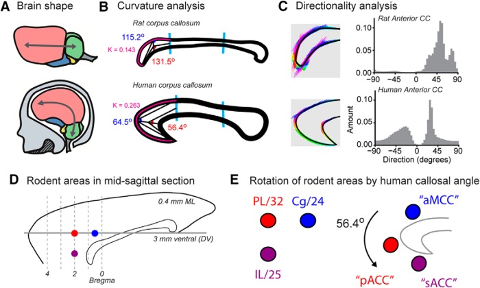
Prefrontal cortex (PFC) means different things to different people. In recent years, there has been a major increase in publications on the PFC, especially using mice. However, inconsistencies in the nomenclature and anatomical boundaries of PFC areas has made it difficult for researchers to compare data and interpret findings across species. We conducted a meta-analysis of publications on the PFC of humans and rodents and found dramatic differences in the focus of research on these species. In addition, we compared anatomical terms and criteria across several common rodent brain atlases and found inconsistencies among, and even within, leading atlases. To assess the impact of these issues on the research community, we conducted a survey of established PFC researchers on their use of anatomical terms and found little consensus. We report on the results of the survey and propose an alternative scheme for interpreting data from rodent studies, based on structural analysis of the corpus callosum and nomenclature used in research on the anterior cingulate cortex (ACC) of primates.

Keywords: atlas; callosum; cingulated; meta-analysis; prefrontal; survey.


Figures

None
Graphical abstract
Figure 1.
Figure 1.
Publication trends in PFC research. A, Publications per year on the PFC of humans, rats, mice, and monkeys from 1945 to 2016. Publication trends on monkey PFC (orange line) have remained unchanged and were not further analyzed. B, Publications on the PFC of mice are appearing at a higher rate than those on rats in the period from 1990 to 2016.
Figure 2.
Figure 2.
Research focus of prefrontal publications. Word clouds with functional color grouping for papers published on the PFC of mice, rats, monkeys, and humans since 2000. Pharmacological terms are purple. Anatomic terms are orange. Terms associated with diseases and other medical conditions are blue. Psychological constructs are green. Other terms are red.
Figure 3.
Figure 3.
Word frequencies across species. A, Hinton plots for anatomic, neurochemical, and disease-related terms in papers focused on the prefrontal cortex of mice, rats, monkeys, and humans. In each plot, columns denote species and rows denote terms. The relative frequency of each term is represented by a square, and the size of the square is defined as the word count divided by total words. Plots are color coded using the same colors as in Figure 2. Most notable, was the use of certain anatomic terms in monkey studies (e.g., FEF, OFC, vlPFC) that are not common in the human or rodent literature. B, Bar plots of the most common terms for rodents (mice and rats) and primates (monkeys and humans). Anatomic terms were sharply divided across orders (rodents versus primates). Rodent studies were focused on the “mPFC,” and primate studies were focused on the “dlPFC.” By contrast, publications focused on neurotransmitters such as dopamine and serotonin were more common in rodent studies, but the relative frequencies of these terms were not discordant across species. A somewhat different finding was apparent in relative word frequencies for diseases and disorders. Rodent studies, especially in rats, more often addressed stress than studies in primates, and primate studies more often addressed schizophrenia compared to the rodent literature.
Figure 4.
Figure 4.
Anatomic terms for rodent PFC areas. A, Sections from the 4th (1998) and 7th (2014) editions of the commonly used Paxinos and Watson rat brain atlas are shown for the most rostral part of the rodent PFC. The 4th edition of the atlas characterized the medial PFC areas as the prelimbic (PrL) and medial orbital (MO) areas. These terms are still used in many recent publications. The 7th edition revised these regions using terms based on Brodmann’s numbers, as dorsal and ventral parts of area 32, but the medial orbital term was retained. B, Sections from the middle parts of the rodent PFC contain more distinct cytoarchitectural areas. Anterior cingulate (Cg1), PrL, and infralimbic (IL) were used in the 4th edition of the atlas. Areas 24 (a and b), 33, and 25 were used in the 7th edition. C, Sections from the most caudal level of the rodent PFC, just anterior to bregma. The areas denoted as Cg1 and Cg2 were relabeled as areas 24a and 24b, respectively, and a new region (to rodent atlases) “area 33” emerged. D, The locations of the sections in A–C are depicted in a midsagittal section. Please note that it was not possible to use atlas sections at the same anterior-posterior locations due to changes in the content of the editions of the atlases. For example, the 4th edition did not include a section at +4.6 AP and the 7th edition did not include a section at +4.2 AP.
Figure 5.
Figure 5.
Crowdsourcing the rodent PFC. A, B, 38 respondents were asked questions about how different cytoarchitectural areas fit into the concept of rodent PFC. Major questions included “Is the prelimbic cortex part of the ACC?,” “Which areas comprise the dorsomedial and ventromedial PFC?,” “Is M2 part of the PFC?,” and if two distinct levels of the medial wall cortex are part of the PFC (located anterior to the genu of the corpus callosum, blue box in B; located below bregma, red box in B). C, Only 8 of 32 (25%) respondents working with rodents said that the prelimbic cortex is part of the ACC. Eight of 20 (40%) respondents working with primates answered this question positively. No differences were apparent in the respondents’ confidence in this answer. Given the small sample size, the difference between rodent and primate researchers was not significant (proportions test: χ2 = 0.69, df = 1, p = 0.4058). This outcome was surprising given that the prelimbic cortex has been considered as a core ACC region (Vogt and Gabriel, 1993). D, There was almost universal agreement that Cg1 is part of the dmPFC (32 of 36 said yes) and IL is part of the vmPFC (33 of 36 said yes). Respondents were divided about how to characterize PL, with 24 of 36 saying it is part of dmPFC and 16 of 36 saying it is part of vmPFC (six respondents included PL in both dmPFC and vmPFC!). E, All respondents agreed that the pregenual region (blue box in B) is part of the rodent PFC. Roughly 25% of respondents felt that the medial wall cortex at bregma (red box in B) and the secondary motor cortex (M2) are PFC regions.
Figure 6.
Figure 6.
Proposed cross-species homologies. A, Rodent brains are flat. Modern primate brains are curved. B, Quantification of the curvature of anterior corpus callosum (using the Kappa library for FIJI). The point of maximum curvature along the superior edge of the anterior third of the corpus callosum is 1.84 times more curved in humans (K = 0.263) than in rats (K = 0.143). Interior angles with vertex at the point of maximum curvature. The rat callosum has obtuse interior angles. The human callosum has acute interior angles. C, Directionality analysis (using FIJI) of the anterior third of the callosum reveals unimodality in rat with a peak of approximately -52°. The human CC peaks at -26° and 16°, indicative of its parabolic shape. A third peak at ∼54° is due to the internal angle of the rostrum. Images used for the directionality analysis are shown on the left and directions are encoded with false color (rainbow colormap with red equal to -90° and violet equal to +90°). D, Locations of three main parts of the rodent PFC (Cg1, blue; PL, red; IL, purple) shown on a midsagittal section (0.4 mm ML). E, Rotation of the three points representing each cortical region by the measured curvature of the human callosum shifts the rodent PFC areas into approximate locations associated with the major divisions of the human medial frontal cortex. This analysis suggests that (1) the area called Cg1 in rodents may be homologous to the anterior part of the midcingulate cortex (aMCC) in primates, (2) the area called PL in rodents may be homologous to the called pACC (pregenual ACC) in primates, and (3) the area called IL in rodents may be homologous to the area called sACC (subgenual ACC) in primates.

References

    1. Aboitiz F, Montiel J (2003) One hundred million years of interhemispheric communication: the history of the corpus callosum. Braz J Med Biol Res 36:409–420. - PubMed
    1. Allen Institute for Brain Science (2008) Technical white paper: Allen Reference Atlas – version 1 (2008) [White Paper] Retrieved from http://help.brain-map.org/display/mousebrain/Documentation 10.1523/ENEURO.0315-18.2018.h - DOI
    1. Amarante LM, Caetano MS, Laubach M (2017) Medial frontal theta is entrained to rewarded actions. J Neurosci 37:10757–10769. - PMC - PubMed
    1. An X, Bandler R, Ongür D, Price JL (1998) Prefrontal cortical projections to longitudinal columns in the midbrain periaqueductal gray in macaque monkeys. J Comp Neurol 401:455–479. - PubMed
    1. Baeg EH, Kim YB, Huh K, Mook-Jung I, Kim HT, Jung MW (2003) Dynamics of population code for working memory in the prefrontal cortex. Neuron 40:177–188. - PubMed