

Impact of endoscopic ultrasonography on diagnosis of pancreatic cancer
- PMID: 30406288
- PMCID: PMC6314985
- DOI: 10.1007/s00535-018-1519-2
Impact of endoscopic ultrasonography on diagnosis of pancreatic cancer
Abstract
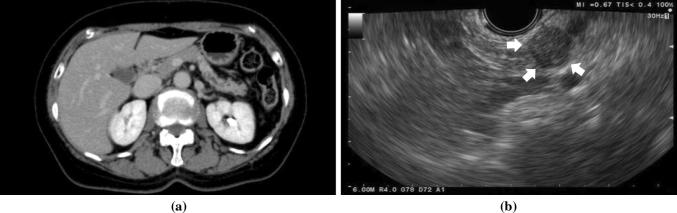
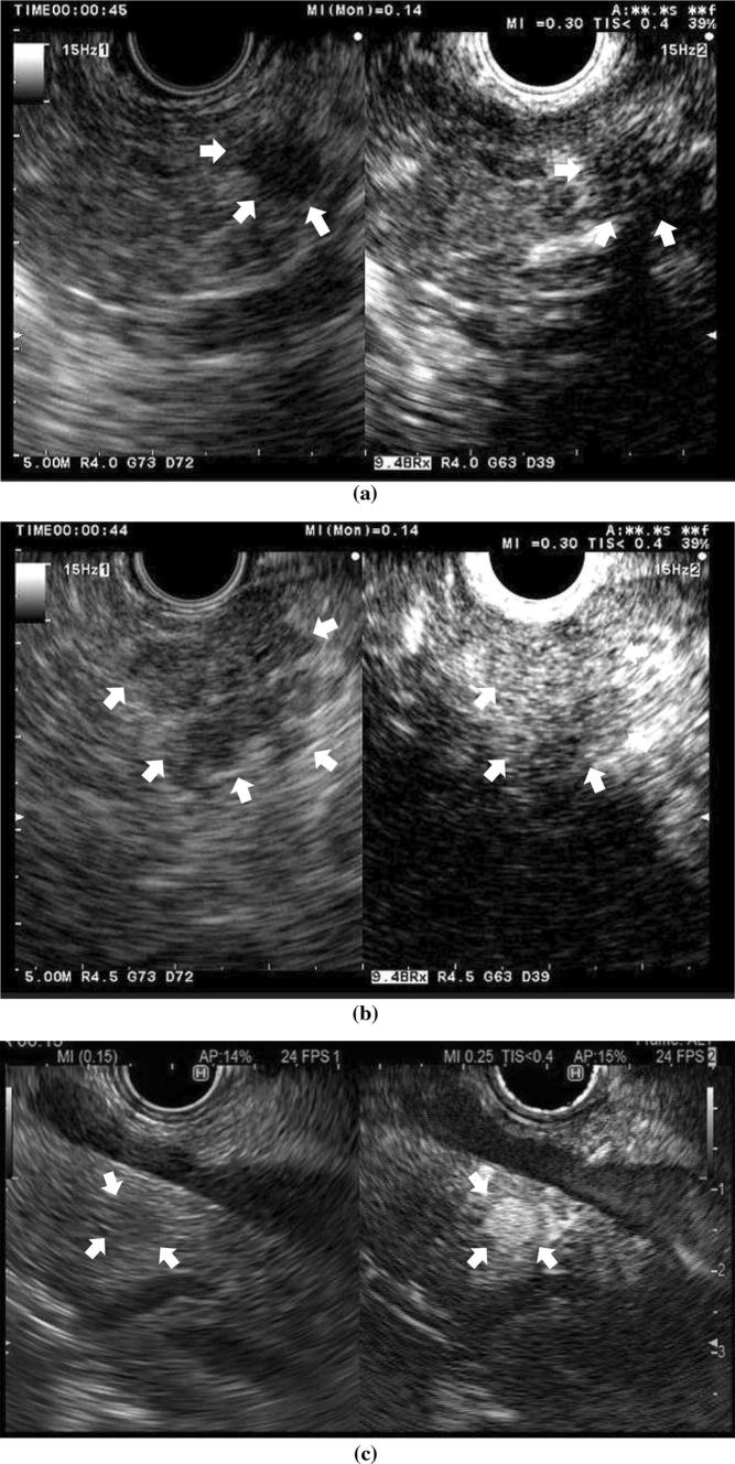
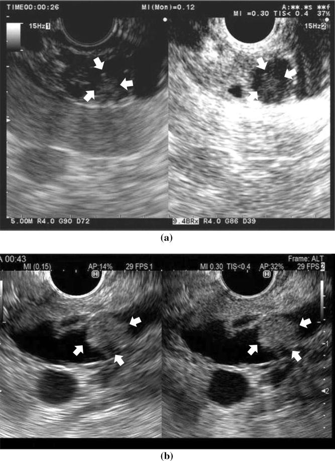
Accumulated evidence has revealed that endoscopic ultrasonography (EUS) has had a great impact on the clinical evaluation of pancreatic cancers. EUS can provide high-resolution images of the pancreas with a quality regarded as far surpassing that achieved on transabdominal ultrasound (US), computed tomography (CT), or magnetic resonance imaging (MRI). EUS is particularly useful for the detection of small pancreatic lesions, while EUS and its related techniques such as contrast-enhanced EUS (CE-EUS), EUS elastography, and EUS-guided fine needle aspiration (EUS-FNA) are also useful in the differential diagnosis of solid or cystic pancreatic lesions and the staging (T-staging, N-staging, and M-staging) of pancreatic cancers. In the diagnosis of pancreatic lesions, CE-EUS and EUS elastography play a complementary role to conventional EUS. When sampling is performed using EUS-FNA, CE-EUS and EUS elastography provide information on the target lesions. Thus, conventional EUS, CE-EUS, EUS elastography, and EUS-FNA are essential in the clinical investigation of pancreatic cancer.
Keywords: Contrast-enhanced endoscopic ultrasonography; Endoscopic ultrasonography; Pancreatic cancer.
Figures
References
-
- The Editorial Board of the Cancer Statistics in Japan. Cancer Registry and Statistics. Cancer Information Service NCCJ (2018) Cancer Statics in Japan 2017. Foundation for Promotion of Cancer Research (FPCR). https://ganjoho.jp/en/professional/statistics/brochure/2017_en.html?.
-
- Noone AM, Howlader N, Krapcho M, Miller D, Brest A, Yu M, Ruhl J, Tatalovich Z, Mariotto A, Lewis DR, Chen HS, Feuer EJ, Cronin KA (eds). SEER cancer statistics review, 1975–2015. National Cancer Institute, Bethesda, MD. Based on November 2017 SEER data. 2018. https://seer.cancer.gov/csr/1975_2015/.
-
- Kato T, Tsukamoto Y, Naitoh Y, et al. Ultrasonographic and endoscopic ultrasonographic angiography in pancreatic mass lesions. Acta Radiol. 1995;36:381–387. - PubMed
-
- Dietrich CF, Ignee A, Frey H. Contrast-enhanced endoscopic ultrasound with low mechanical index: a new technique. Z Gastroenterol. 2005;43:1219–1223. - PubMed
-
- Quaia E. Classification and safety of microbubble-based contrast agents. In: Quaia E, editor. Contrast media ultrason. New York: Springer; 2005. pp. 3–14.
Publication types
MeSH terms
LinkOut - more resources
Full Text Sources
Medical
