

Pneumocardia and septic pulmonary embolism due to nongas-forming liver abscess: A case report
- PMID: 30407318
- PMCID: PMC6250546
- DOI: 10.1097/MD.0000000000013096
Pneumocardia and septic pulmonary embolism due to nongas-forming liver abscess: A case report
Abstract
Rationale: Pneumocardia and septic pulmonary embolism are uncommon complications of Klebsiella pneumoniae primary liver abscess (KPLA); however, they may lead to a poor clinical outcome.
Patient concerns: A 67-year-old woman was admitted to our hospital with fever, chills, cough, and dyspnea for 4 days. She had a previous history of diabetes mellitus.
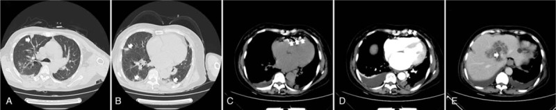
Diagnoses: The chest computed tomography (CT) revealed multiple peripheral nodules in both lungs and wedge-shaped peripheral infiltrative lesions abutting the pleura, suggestive of septic pulmonary embolism. An abdominal CT on the following day showed a large liver abscess without gas formation and pneumocardia of the right ventricle.
Interventions: After the antibiotic therapy of intravenous imipenem and drainage of the liver abscess, our patient made a complete recovery.
Outcomes: The patient was discharged on the 25th hospital day after full recovery and was doing well on follow-up at 10 months.
Lessons: KPLA is potentially fatal due to the associated serious metastatic complications. Attention must be paid not only to the primary focus of infection but also to infection of other organs. It is important to detect to diagnose the spread of infection accurately, in a timely manner, to improve the prognosis of this condition.
Conflict of interest statement
The authors have no conflicts of interest.
Figures
Similar articles
-
A CARE-compliant article: a case report of pleural empyema secondary to Klebsiella pneumoniae liver abscess with a hepatopleural fistula.Medicine (Baltimore). 2020 Apr;99(16):e19869. doi: 10.1097/MD.0000000000019869. Medicine (Baltimore). 2020. PMID: 32312012 Free PMC article.
-
Septic pulmonary embolism caused by a Klebsiella pneumoniae liver abscess: clinical characteristics, imaging findings, and clinical courses.Clinics (Sao Paulo). 2015 Jun;70(6):400-7. doi: 10.6061/clinics/2015(06)03. Epub 2015 Jun 1. Clinics (Sao Paulo). 2015. PMID: 26106957 Free PMC article.
-
Septic pulmonary emboli secondary to pyogenic liver abscess in a diabetic patient.Intern Med. 1995 Jan;34(1):42-5. doi: 10.2169/internalmedicine.34.42. Intern Med. 1995. PMID: 7718979
-
Emphysematous liver abscesses complicated by septic pulmonary emboli in patients with diabetes: two cases.Intern Med. 2013;52(1):141-5. doi: 10.2169/internalmedicine.52.8737. Epub 2013 Jan 1. Intern Med. 2013. PMID: 23291690 Review.
-
Liver abscess in the caudate lobe caused by Klebsiella pneumoniae: a rare case report and literature review.BMC Infect Dis. 2024 Jul 19;24(1):708. doi: 10.1186/s12879-024-09569-6. BMC Infect Dis. 2024. PMID: 39030483 Free PMC article. Review.
Cited by
-
Endogenous Klebsiella endophthalmitis as the presentation of both Klebsiella liver abscess and underlying anti-IFN-3 autoimmunity.Access Microbiol. 2020 Aug 28;2(11):acmi000164. doi: 10.1099/acmi.0.000164. eCollection 2020. Access Microbiol. 2020. PMID: 33294768 Free PMC article.
References
-
- Sakpal SV, Tichauer M, Chamberlain RS. Hepatic venous gas and pneumocardia. Dig Liver Dis 2009;41:915–6. - PubMed
-
- Oh HG, Cha SI, Shin KM, et al. Risk factors for mortality in patients with septic pulmonary embolism. J Infect Chemother 2016;22:553–8. - PubMed
-
- Keng LT, Fu YH, Lin YH. Gas-forming liver abscess with pneumocardia. J Emerg Med 2014;46:e185–186. doi: 10.1016/j.jemermed.2014.01.021. - PubMed
Publication types
MeSH terms
Substances
LinkOut - more resources
Full Text Sources
Medical
