

Dimensional accuracy of cone beam CT with varying angulation of the jaw to the X-ray beam
- PMID: 30407848
- PMCID: PMC6592582
- DOI: 10.1259/dmfr.20180319
Dimensional accuracy of cone beam CT with varying angulation of the jaw to the X-ray beam
Abstract
Objectives: Cone beam CT (CBCT) machines do not always allow for patients to be scanned in the ideal position for image acquisition. This study aimed to investigate the influence of the position/angulation of the mandible relative to the X-ray beam of a CBCT machine.
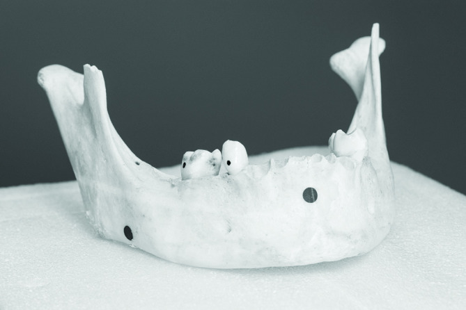
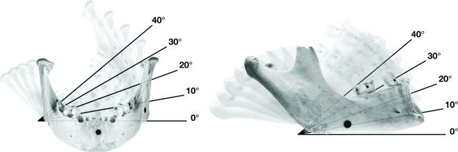
Methods: Five sequential CBCT scans were captured of a human mandible at each angulation of 10°, 20°, 30°, and 40° using a coronal and sagittal positioning. Inspection software utilized a best-fit alignment to automatically calculate the three-dimensional variation at 15 standardized points of interest.
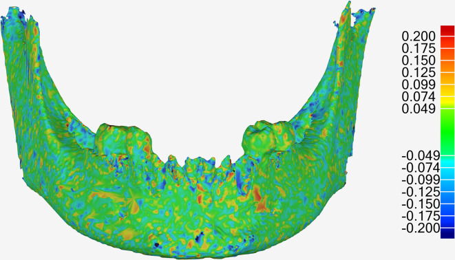
Results: Statistically significant differences were found between the dimensional accuracy of CBCT scans taken at 10° (26.3 µm) of coronal angulation, as well as those taken at 20° (-17.3 mm) and 30° (35.2 mm) of sagittal angulations (p < 0.001, 0.016, and <0.001, respectively). The largest deviations in accuracy included an overall maximum deviation of 490 mm.
Conclusions: The position of the mandible with respect to the X-ray beam has a clinically insignificant effect on dimensional accuracy, up to the maximum angle of 40° assessed.
Keywords: cone beam CT; dimensional measurement accuracy; imaging; three-dimensional.
Figures
Similar articles
-
A comparative evaluation of cone beam CT and micro-CT on trabecular bone structures in the human mandible.Dentomaxillofac Radiol. 2013;42(8):20130145. doi: 10.1259/dmfr.20130145. Epub 2013 Jul 5. Dentomaxillofac Radiol. 2013. PMID: 23833320 Free PMC article.
-
Accuracy and reliability of different cone beam computed tomography (CBCT) devices for structural analysis of alveolar bone in comparison with multislice CT and micro-CT.Eur J Oral Implantol. 2017;10(1):95-105. Eur J Oral Implantol. 2017. PMID: 28327698
-
The impact of different cone beam computed tomography and multi-slice computed tomography scan parameters on virtual three-dimensional model accuracy using a highly precise ex vivo evaluation method.J Craniomaxillofac Surg. 2016 May;44(5):632-6. doi: 10.1016/j.jcms.2016.02.005. Epub 2016 Feb 13. J Craniomaxillofac Surg. 2016. PMID: 27017101
-
Variations in head tilt during the acquisition of cone beam computed tomography scans and their effects on effective radiation dose.Dentomaxillofac Radiol. 2024 Nov 1;53(8):566-572. doi: 10.1093/dmfr/twae043. Dentomaxillofac Radiol. 2024. PMID: 39133160
-
Dimensional measurement accuracy of 3-dimensional models from cone beam computed tomography using different voxel sizes.Oral Surg Oral Med Oral Pathol Oral Radiol. 2021 Sep;132(3):361-369. doi: 10.1016/j.oooo.2021.05.009. Epub 2021 May 30. Oral Surg Oral Med Oral Pathol Oral Radiol. 2021. PMID: 34246615
Cited by
-
The Effect of Mandibular Angulation on Preoperative Assessment of Dental Implant Insertion at Premolar Region: CBCT Study.Biomed Res Int. 2022 May 28;2022:7879239. doi: 10.1155/2022/7879239. eCollection 2022. Biomed Res Int. 2022. PMID: 35669722 Free PMC article.
