

Synaptic distributions of pS214-tau in rhesus monkey prefrontal cortex are associated with spine density, but not with cognitive decline
- PMID: 30408169
- PMCID: PMC6333519
- DOI: 10.1002/cne.24576
Synaptic distributions of pS214-tau in rhesus monkey prefrontal cortex are associated with spine density, but not with cognitive decline
Abstract
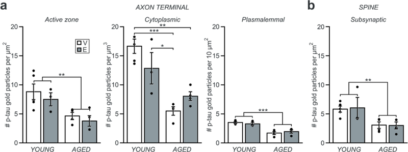
Female rhesus monkeys and women are subject to age- and menopause-related deficits in working memory, an executive function mediated by the dorsolateral prefrontal cortex (dlPFC). Long-term cyclic administration of 17β-estradiol improves working memory, and restores highly plastic axospinous synapses within layer III dlPFC of aged ovariectomized monkeys. In this study, we tested the hypothesis that synaptic distributions of tau protein phosphorylated at serine 214 (pS214-tau) are altered with age or estradiol treatment, and couple to working memory performance. First, ovariectormized young and aged monkeys received vehicle or estradiol treatment, and were tested on the delayed response (DR) test of working memory. Serial section electron microscopic immunocytochemistry was then performed to quantitatively assess the subcellular synaptic distributions of pS214-tau. Overall, the majority of synapses contained pS214-tau immunogold particles, which were predominantly localized to the cytoplasm of axon terminals. pS214-tau was also abundant within synaptic and cytoplasmic domains of dendritic spines. The density of pS214-tau immunogold within the active zone, cytoplasmic, and plasmalemmal domains of axon terminals, and subjacent to the postsynaptic density within the subsynaptic domains of dendritic spines, were each reduced with age. None of the variables examined were directly linked to cognitive status, but a high density of pS214-tau immunogold particles within presynaptic cytoplasmic and plasmalemmal domains, and within postsynaptic subsynaptic and plasmalemmal domains, accompanied high synapse density. Together, these data support a possible physiological, rather than pathological, role for pS214-tau in the modulation of synaptic morphology in monkey dlPFC.
Keywords: RRID:AB_2716719; RRID:AB_2750560; RRID:SCR_002865; RRID:SCR_014199; aging; area 46; delayed response; estradiol; menopause; pS214.
© 2018 Wiley Periodicals, Inc.
Conflict of interest statement
Figures


Similar articles
-
Diverse Synaptic Distributions of G Protein-coupled Estrogen Receptor 1 in Monkey Prefrontal Cortex with Aging and Menopause.Cereb Cortex. 2017 Mar 1;27(3):2022-2033. doi: 10.1093/cercor/bhw050. Cereb Cortex. 2017. PMID: 26941383 Free PMC article.
-
Estrogen Restores Multisynaptic Boutons in the Dorsolateral Prefrontal Cortex while Promoting Working Memory in Aged Rhesus Monkeys.J Neurosci. 2016 Jan 20;36(3):901-10. doi: 10.1523/JNEUROSCI.3480-13.2016. J Neurosci. 2016. PMID: 26791219 Free PMC article.
-
Estrogen Alters the Synaptic Distribution of Phospho-GluN2B in the Dorsolateral Prefrontal Cortex While Promoting Working Memory in Aged Rhesus Monkeys.Neuroscience. 2018 Dec 1;394:303-315. doi: 10.1016/j.neuroscience.2018.09.021. Neuroscience. 2018. PMID: 30482274 Free PMC article.
-
Interactive effects of age and estrogen on cortical neurons: implications for cognitive aging.Neuroscience. 2011 Sep 15;191:148-58. doi: 10.1016/j.neuroscience.2011.05.045. Epub 2011 Jun 2. Neuroscience. 2011. PMID: 21664255 Free PMC article. Review.
-
Neuronal and morphological bases of cognitive decline in aged rhesus monkeys.Age (Dordr). 2012 Oct;34(5):1051-73. doi: 10.1007/s11357-011-9278-5. Epub 2011 Jun 28. Age (Dordr). 2012. PMID: 21710198 Free PMC article. Review.
Cited by
-
Alzheimer's-like pathology in aging rhesus macaques: Unique opportunity to study the etiology and treatment of Alzheimer's disease.Proc Natl Acad Sci U S A. 2019 Dec 26;116(52):26230-26238. doi: 10.1073/pnas.1903671116. Epub 2019 Dec 23. Proc Natl Acad Sci U S A. 2019. PMID: 31871209 Free PMC article.
-
Tau and tauopathies across primate species: implications for modeling neurodegenerative disorders.Front Aging Neurosci. 2025 Jul 23;17:1598245. doi: 10.3389/fnagi.2025.1598245. eCollection 2025. Front Aging Neurosci. 2025. PMID: 40771194 Free PMC article. Review.
-
Comparative neuropathology in aging primates: A perspective.Am J Primatol. 2021 Nov;83(11):e23299. doi: 10.1002/ajp.23299. Epub 2021 Jul 13. Am J Primatol. 2021. PMID: 34255875 Free PMC article. Review.
References
-
- Alonso AD, Grundke-Iqbal I, Barra HS, & Iqbal K (1997). Abnormal phosphorylation of tau and the mechanism of Alzheimer neurofibrillary degeneration: sequestration of microtubule-associated proteins 1 and 2 and the disassembly of microtubules by the abnormal tau. Proc Natl Acad Sci U S A,94(1), 298–303. - PMC - PubMed
-
- Andersen K, Launer LJ, Dewey ME, Letenneur L, Ott A, Copeland JR, … Hofman A (1999). Gender differences in the incidence of AD and vascular dementia: The EURODEM Studies. EURODEM Incidence Research Group. Neurology, 53(9), 1992–1997. - PubMed
