. 2019 Jul;1866(7):1151-1161.
doi: 10.1016/j.bbamcr.2018.10.022. Epub 2018 Nov 5.

Mining of Ebola virus entry inhibitors identifies approved drugs as two-pore channel pore blockers

Affiliations

Mining of Ebola virus entry inhibitors identifies approved drugs as two-pore channel pore blockers

Christopher J Penny et al. Biochim Biophys Acta Mol Cell Res. 2019 Jul.

Abstract

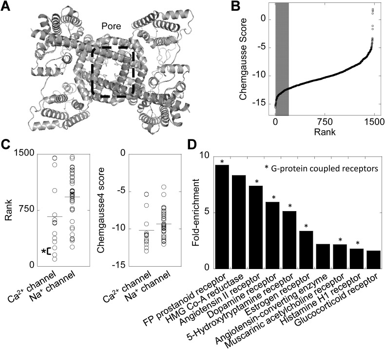
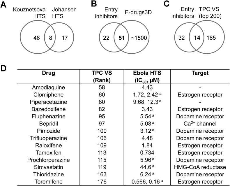
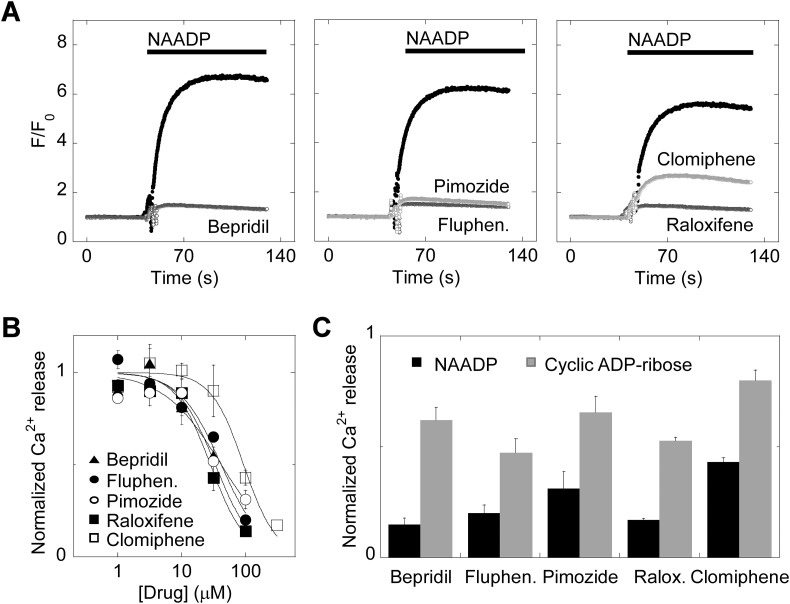
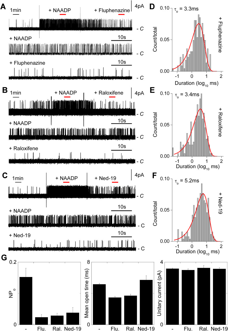
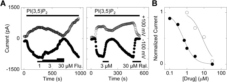
Two-pore channels (TPCs) are Ca2+-permeable ion channels localised to the endo-lysosomal system where they regulate trafficking of various cargoes including viruses. As a result, TPCs are emerging as important drug targets. However, their pharmacology is ill-defined. There are no approved drugs to target them. And their mechanism of ligand activation is largely unknown. Here, we identify a number of FDA-approved drugs as TPC pore blockers. Using a model of the pore of human TPC2 based on recent structures of mammalian TPCs, we virtually screened a database of ~1500 approved drugs. Because TPCs have recently emerged as novel host factors for Ebola virus entry, we reasoned that Ebola virus entry inhibitors may exert their effects through inhibition of TPCs. Cross-referencing hits from the TPC virtual screen with two recent high throughput anti-Ebola screens yielded approved drugs targeting dopamine and estrogen receptors as common hits. These compounds inhibited endogenous NAADP-evoked Ca2+ release from sea urchin egg homogenates, NAADP-mediated channel activity of TPC2 re-routed to the plasma membrane, and PI(3,5)P2-mediated channel activity of TPC2 expressed in enlarged lysosomes. Mechanistically, single channel analyses showed that the drugs reduced mean open time consistent with a direct action on the pore. Functionally, drug potency in blocking TPC2 activity correlated with inhibition of Ebola virus-like particle entry. Our results expand TPC pharmacology through the identification of approved drugs as novel blockers, support a role for TPCs in Ebola virus entry, and provide insight into the mechanisms underlying channel regulation. This article is part of a Special Issue entitled: ECS Meeting edited by Claus Heizmann, Joachim Krebs and Jacques Haiech.

Keywords: Ca(2+); Ebola virus; Lysosomes; NAADP; TPC2; Virtual screening.


Figures

Fig. 1
Fig. 1
A structure-based virtual screen of approved drugs based on the TPC2 pore. A, structural model of human TPC2 viewed from the cytosolic face. The pore region is highlighted by the dashed box. B, results of a virtual screen of the e-Drugs3D database against the pore region of TPC2. The top 200 drugs are marked by the shaded region. C, plots showing the rank of individual drugs from the virtual screen targeting voltage-gated Ca2+ and Na+ channels. The horizontal lines represent the mean. Nicardipine (rank #154) and diltiazem (rank #247) are highlighted*. D, enrichment analysis of drug classes within the top 200 hits. G-protein coupled receptors are highlighted*.
Fig. 2
Fig. 2
Identification of drugs targeting dopamine and estrogen receptors as putative TPC2 inhibitors. A, Venn diagram depicting the relationship between approved drugs identified as Ebola virus entry inhibitors in independent high through put screens by Kouznetsova [58] and Johansen et al. [59]. B, Venn diagram depicting the relationships between the entry inhibitors identified in A with the e-Drugs3D database used for the TPC virtual screen. C, Venn diagram depicting the relationship between entry inhibitors present in the e-Drugs3-D database and the top 200 hits from the TPC virtual screen. D, prioritised set of 14 drugs identified from C listing their rank in the TPC virtual screen, their reported IC50 values for inhibiting virus entry (from Kouznetsova [58] and/or aJohansen et al. [59]) and their primary target (annotated in e-Drugs3D).
Fig. 3
Fig. 3
Ca2+ channel blockers, dopamine antagonists and selective estrogen receptor modulators block endogenous NAADP-mediated Ca2+ signals. A, exemplar Ca2+ signals, recorded using Fluo-4 from sea urchin egg homogenates pre-incubated with the indicated drug (100 μM) or DMSO (black traces) for ~1 min prior to stimulation with NAADP (1 μM). B, concentration-effect relationships for blockade of NAADP responses by the various drugs (n = 3). Data are normalised to the NAADP response in the presence of DMSO. C, summary data quantifying the effect of the drugs (100 μM) on Ca2+ response to NAADP versus cyclic ADP-ribose (n = 3). Data are normalised to the NAADP and cyclic ADP-ribose responses in the presence of DMSO.
Fig. 4
Fig. 4
NAADP stimulates single channel activity of TPC2 rerouted to the plasma membrane. A, exemplar single channel recordings from inside-out patches excised from HEK cells expressing plasma membrane-targeted TPC2 in response to the indicated concentrations of NAADP. Na+ was the charge carrier and the holding potential was +40 mV. C denotes the closed state. B, summary data quantifying open probability (NPo; n = 3–28). C, current-voltage relationships derived from records similar to those shown in A (n = 3). D, exemplar open and closed time distributions at the indicated NAADP concentration. E, summary data quantifying mean open time (n = 3–28). F, likely gating schemes for TPC2.
Fig. 5
Fig. 5
Dopamine antagonists and selective estrogen receptor modulators inhibit single channel activity of TPC2 by reducing open time. A–C, exemplar single channel records from inside-out patches expressing plasma membrane-targeted TPC2. Patches were sequentially challenged with 100 nM NAADP and either fluphenazine (100 μM; A), raloxifene (100 μM; B) or Ned-19 (1 μM; C). Large vertical deflections are addition artefacts. Red bars indicate the sections of the upper trace in the presence of NAADP before and after drug addition that were expanded in the corresponding lower traces. D–F, exemplar open time distributions from records similar to A–C in the presence of the indicated drug. G, summary data quantifying open probability, mean closed time and unitary conductance for TPC2 activity in response to 100 nM NAADP in the absence (n = 10) and presence of the indicated drug (n = 3–4). (For interpretation of the references to color in this figure legend, the reader is referred to the web version of this article.)
Fig. 6
Fig. 6
Dopamine antagonists and selective estrogen receptor modulators inhibit lysosomal TPC2 currents. A, exemplar currents recorded from enlarged lysosomes isolated from HEK cells expressing lysosome-targeted TPC2 in response to PI(3,5)P2 (1 μM). The holding potentials were +100 mV (upper traces) and −100 mV (lower traces). Lysosomes were challenged with fluphenazine (Flu.) and raloxifene (Ral.) at the concentrations and for the times indicated. B, concentration-effect relationships for blockade of PI(3,5)P2 currents by fluphenazine (open circles) and raloxifene (closed circles;n = 2). Data are normalised to the PI(3,5)P2 response in the absence of the drugs.
Fig. 7
Fig. 7
Dopamine antagonists and selective estrogen receptor modulators inhibit entry of Ebola virus-like particles. A, infection of HeLa cells with Ebola virus-like particles in the absence and presence of EIPA (11.1 μM; n = 3) or tetrandrine (1.2 μM; n = 4). B, concentration-effect relationships for blockade of virus-like particle entry by the indicated dopamine antagonist (squares) or SERMs (circles). n = 3–4.
Fig. S1
Fig. S1
Current amplitude histograms of single channel records from inside-out patches expressing plasma membrane-targeted TPC2. A–C, Histograms in the absence or presence of the indicated concentration of NAADP. D–E, Histograms in the presence NAADP (100 nM) and the indicated drug.

Similar articles

Cited by

References

    1. Churchill G.C., Okada Y., Thomas J.M., Genazzani A.A., Patel S., Galione A. NAADP mobilizes Ca2+ from reserve granules, lysosome-related organelles, in sea urchin eggs. Cell. 2002;111:703–708. - PubMed
    1. Lee H.C. NAADP-mediated calcium signaling. J. Biol. Chem. 2005;280(40):33693–33696. - PubMed
    1. Patel S., Docampo R. Acidic calcium stores open for business: expanding the potential for intracellular Ca2+ signaling. Trends Cell Biol. 2010;20:277–286. - PMC - PubMed
    1. Morgan A.J., Platt F.M., Lloyd-Evans E., Galione A. Molecular mechanisms of endolysosomal Ca2+ signalling in health and disease. Biochem. J. 2011;439:349–374. - PubMed
    1. Patel S., Cai X. Evolution of acid Ca2+ stores and their resident Ca2+-permeable channels. Cell Calcium. 2015;57:222–230. - PubMed

Publication types