. 2019;20(1):42-51.
doi: 10.1080/15384047.2018.1504722. Epub 2018 Nov 8.

Caprine herpesvirus 1 (CpHV-1) as a potential candidate for oncolytic virotherapy

Affiliations

Caprine herpesvirus 1 (CpHV-1) as a potential candidate for oncolytic virotherapy

Serena Montagnaro et al. Cancer Biol Ther. 2019.

Abstract

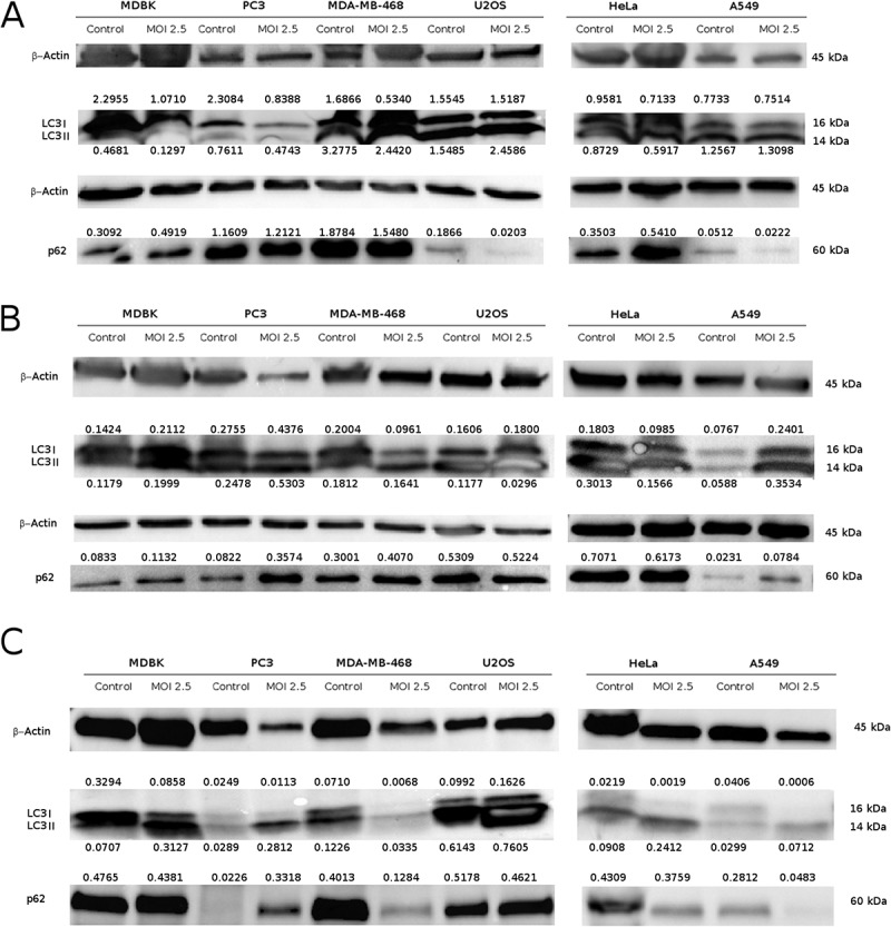
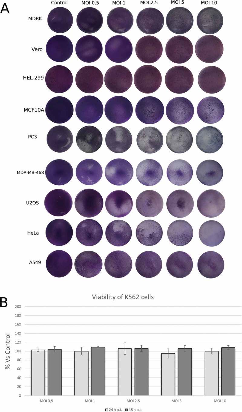
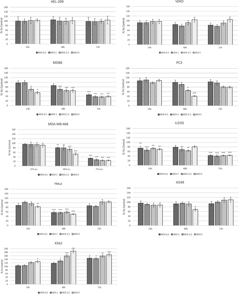
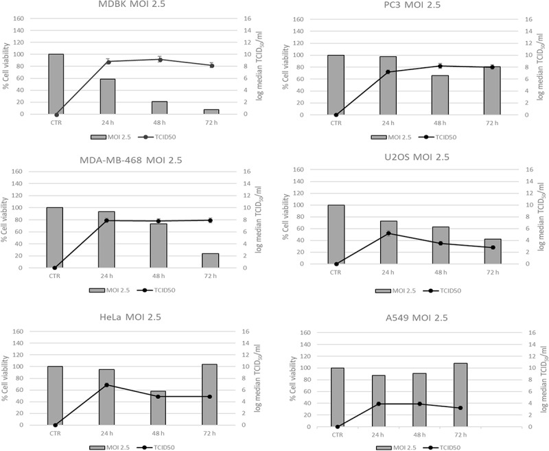
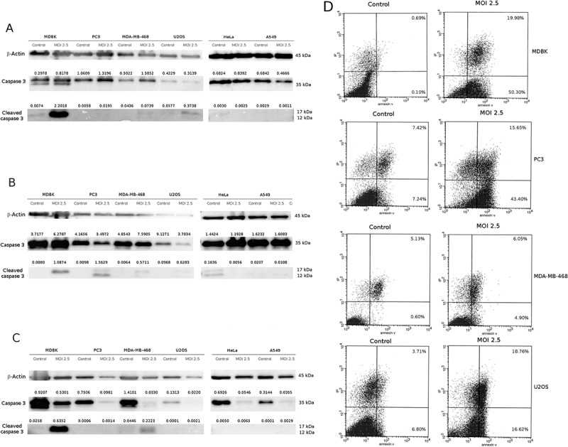
Caprine Herpesvirus type 1 (CpHV-1) is a species-specific herpes virus able to induce apoptosis in several biological systems. In the present study we aimed to investigate the ability of CpHV-1 to reduce cells viability, to replicate and to cause cell death also in human cancer cell lines. We tested the CpHV-1 effects on HEL-299, Vero, MDA-MB-468, HeLa, U2OS, PC3, A549 and K562 neoplastic cell lines and on MDBK cells. Firstly, we evaluated the effect of CpHV-1 infection on cell viability by MTT assay and our data showed that CpHV-1 can induce a marked cytopathic effect (CPE) in most of cell lines tested, except for HEL-299, Vero and K562 cells. The reduction of cell viability was associated with a significant increase of viral production. We next investigated if CpHV-1 was able to induce cell death and so through western blotting analysis we evaluated cleaved caspase 3, LC3II and p62 protein levels after infection. Caspase 3 activation was detected in MDBK cells and, even if at different times p.i., also in MDA-MB-468, U2OS, and PC3 cell lines, while LC3II increase and concomitant p62 protein reduction were observed only in U2OS, and A549 cells, no significant alteration of these proteins was observed in the other cell lines tested. Finally, to confirm virus ability to trigger apoptosis we performed an Annexin-V apoptosis test after 24 h p.i. Although we need to further explore mechanisms underlying CpHV-1 treatment, this study could serve as the basis for the development of new treatment options aiming to fight several cancer types.

Keywords: Oncolytic virotherapy; apoptosis; autophagy; human cancer cells.


Figures

Figure 5.
Figure 5.
Evaluation of autophagy markers in neoplastic cell lines after CpHV-1 infection. Western blotting analysis of LC3I, LC3II and p62 levels in MDBK, PC3, MDA-MB-468, U2OS, Hela and A549 cells were performed after mock infection or infection with CpHV-1 at MOI 2.5, at 12 h (panel A), 24 h (panel B) and 48 h (panel C) p.i.. β-Actin were used as a loading control. Band densitometry values indicate LC3I, LC3II and p62 levels normalized to β-Actin levels.
Figure 1.
Figure 1.
Permissiveness of human neoplastic cell lines to Caprine Herpesvirus 1 (CpHV-1). Cells were infected with the indicated MOI of CpHV-1 at 24 h post infection and stained with Giemsa to observe Cytopathic effect (CPE). Panel B show viability results in cell line determined by trypan blue dye exclusion method at 24 h and 48 h p.i. Results are expressed as the mean ± SD of three independent experiments performed in duplicate. The SD was calculated on the raw data and expressed as a percentage.
Figure 2.
Figure 2.
Dose–response curve of human cancer cell lines infected with different MOI of CpHV-1 at different times p.i.. MTT test was performed at different hours post infection and the absorbance assayed as described in the Materials and Methods Section. Data are presented as a percentage of the cell viability calculated respect to control and results are expressed as the mean ± SD of three independent experiments performed in duplicate. Statistical differences between control and CpHV-1 infected groups were evaluated by one-way analysis of Variance (ANOVA), followed by Turkey’s post-test and indicated by probability P. *P < 0.05, **P < 0.01, and ***P < 0.001. The SD was calculated on the raw data and expressed as a percentage.
Figure 3.
Figure 3.
Time course of virus production in Human cancer cell lines infected with CpHV-1. Virus production was titrated at 24 h, 48 h and 72 h after infection with CpHV-1 at a MOI. of 2.5. Results are expressed as Mean ± SD of three independent experiments performed in duplicate.
Figure 4.
Figure 4.
CpHV-1 infection induces apoptosis in neoplastic cell lines. Panel A-C: Western blotting analysis of Caspase and Cleaved caspase 3 levels in MDBK, PC3, MDA-MB-468, U2OS, Hela and A549 cells were performed after mock infection or infection with CpHV-1 at MOI 2.5, at 12 h (panel A), 24 h (panel B) and 48 h (panel C) p.i.. β-Actin were used as a loading control. Band densitometry values indicate caspase 3 and cleaved caspase 3 levels normalized to β-Actin levels. Panel D: Annexin V apoptosis analysis. MDBK, PC3, MDA-MB-468 and U2Os cells were infected with CpHV-1 virus and after 24 h analyzed by annexin-V assay. The graphs (panel D) show the percentages of early apoptosis (population of cells positive for annexin-V staining) and late apoptosis/necrosis (population of cells positive for both annexin-V and propidium iodide staining).

Similar articles

Cited by

References

    1. Fountzilas C, Patel S, Mahalingam D.. Oncolytic virotherapy, updates and future directions. Oncotarget. 2017;8(60):102617–102639. doi:10.18632/oncotarget.18309. - DOI - PMC - PubMed
    1. Rodrigues R, Cuddington B, Mossman K. Bovine herpesvirus type 1 as a novel oncolytic virus. Cancer Gene Ther. 2010;17:344–355. doi:10.1038/cgt.2009.77. - DOI - PubMed
    1. Courchesne MJ, White MC, Stanfield BA, Frampton AR Jr.. Equine herpesvirus type 1-mediated oncolysis of human glioblastoma multiforme cells. J Virol. 2012;86(5):2882–2886. doi:10.1128/JVI.06296-11. - DOI - PMC - PubMed
    1. Kaufman HL, Kohlhapp FJ, Zloza A. Oncolytic viruses: a new class of immunotherapy drugs. Nat Rev Drug Discov. 2016;14(9):642–662. doi:10.1038/nrd4663. - DOI - PMC - PubMed
    1. Andtbacka RH, Kaufman HL, Collichio F, Amatruda T, Senzer N, Chesney J, Delman KA, Spitler LE, Puzanov I, Agarwala SS, et al. Talimogene laherparepvec improves durable response rate in patients with advanced melanoma. J Clin Oncol. 2015;33:2780–2788. doi:10.1200/JCO.2014.58.3377. - DOI - PubMed

Publication types