

Relevance of vitamin D deficiency in patients with chronic autoimmune atrophic gastritis: a prospective study
- PMID: 30409113
- PMCID: PMC6225568
- DOI: 10.1186/s12876-018-0901-0
Relevance of vitamin D deficiency in patients with chronic autoimmune atrophic gastritis: a prospective study
Abstract
Background: Chronic autoimmune atrophic gastritis (CAAG) is an autoimmune disease characterized by hypo/achlorhydria. A role of CAAG in the pathogenesis of nutritional deficiencies has been reported, therefore we hypothesized a possible association between CAAG and 25-OH-Vitamin D [25(OH)D] deficiency. Aim of the present study is to evaluate the prevalence of 25(OH)D deficiency in CAAG patients.
Methods: 87 CAAG patients (71 females; mean age 63.5 ± 12.8 years) followed at our Centre from January 2012 to July 2015 were consecutively evaluated. 25(OH)D, vitamin B12, parathormone, and calcium were measured in all the CAAG patients. The results were compared with a control group of 1232 healthy subjects.
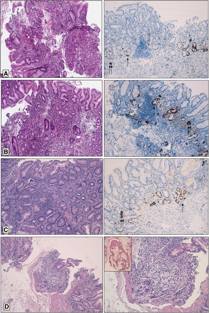
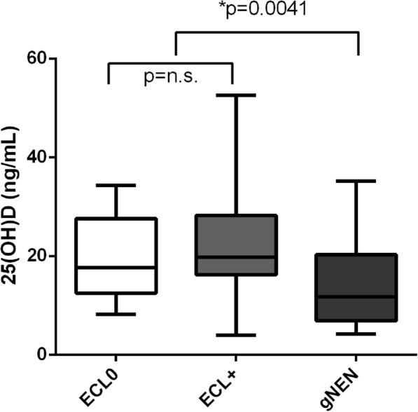
Results: In the CAAG group the mean 25(OH)D levels were significantly lower than in the control group (18.8 vs. 27.0 ng/ml, p < 0.0001). 25(OH)D levels < 20 ng/ml was observed in 57 patients, while levels < 12.5 ng/ml in 27 patients. A significant correlation between vitamin B12 values at diagnosis and 25(OH)D levels was observed (rs = 0.25, p = 0.01). Interestingly, the CAAG patients with moderate/severe gastric atrophy had lower 25(OH)D values as compared to those with mild atrophy (11.8 vs. 20 ng/ml; p = 0.0047). Moreover, the 25(OH)D levels were significantly lower in CAAG patients with gastric carcinoid as compared to those without gastric carcinoid (11.8 vs. 19.8 ng/ml; p = 0,0041).
Conclusion: Data from the present study showed a significant reduction of 25(OH)D levels in CAAG patients and a possible impairment of vitamin D absorption in CAAG may be postulated. Any implication to the genesis of gastric carcinoids remains to be elucidated.
Keywords: Bone health; Chronic autoimmune atrophic gastritis; Gastric carcinoid; Osteoporosis; Vitamin D deficiency.
Conflict of interest statement
Ethics approval and consent to participate
All the subjects, after full explanation of the purpose and nature of all procedures used, gave their written informed consent to participate in the study, which was approved by the Ethics Committee of the Fondazione IRCCS Ca′ Granda Ospedale Maggiore Policlinico of Milan.
Consent for publication
Not Applicable.
Competing interests
The authors declare that there is no conflict of interest that could be perceived as prejudicing the impartiality of the research reported
Publisher’s Note
Springer Nature remains neutral with regard to jurisdictional claims in published maps and institutional affiliations.
Figures
References
-
- Solcia E, Capella C, Fiocca R, Cornaggia M, Rindi G, Villani L, et al. Exocrine and endocrine epithelial changes in type A and B chronic gastritis. In: Malfertheiner P, Ditschuneit H, et al., editors. Helicobacter pylori, Gastritis and Peptic Ulcer. Berlin: Springer Berlin Heidelberg; 1990. pp. 245–258.
-
- Wintrobe M, Lee G, Boggs D, Bithell T, Foerster J, Athens J, et al. et al. Megaloblastic and nonmegaloblastic macrocytic anemias. In: Wintrobe M, Lee G, Boggs D, Bithell T, Foerster J, et al.et al., editors. Clinical hematology. 8. Philadelphia: Lea & Febiger; 1981. pp. 559–604.
MeSH terms
Substances
LinkOut - more resources
Full Text Sources
Medical
