. 2018 Nov 9;11(1):64.
doi: 10.1186/s13041-018-0408-1.

Exposure to mild blast forces induces neuropathological effects, neurophysiological deficits and biochemical changes

Affiliations

Exposure to mild blast forces induces neuropathological effects, neurophysiological deficits and biochemical changes

Adan Hernandez et al. Mol Brain. .

Erratum in

Abstract

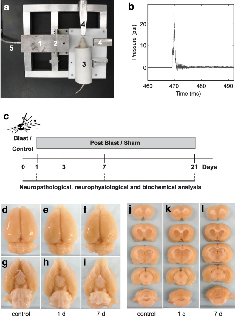
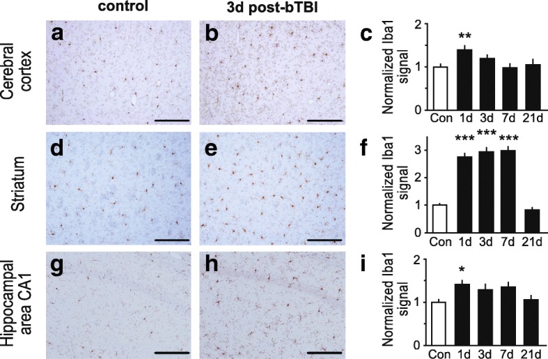
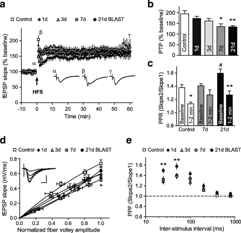
Direct or indirect exposure to an explosion can induce traumatic brain injury (TBI) of various severity levels. Primary TBI from blast exposure is commonly characterized by internal injuries, such as vascular damage, neuronal injury, and contusion, without external injuries. Current animal models of blast-induced TBI (bTBI) have helped to understand the deleterious effects of moderate to severe blast forces. However, the neurological effects of mild blast forces remain poorly characterized. Here, we investigated the effects caused by mild blast forces combining neuropathological, histological, biochemical and neurophysiological analysis. For this purpose, we employed a rodent blast TBI model with blast forces below the level that causes macroscopic neuropathological changes. We found that mild blast forces induced neuroinflammation in cerebral cortex, striatum and hippocampus. Moreover, mild blast triggered microvascular damage and axonal injury. Furthermore, mild blast caused deficits in hippocampal short-term plasticity and synaptic excitability, but no impairments in long-term potentiation. Finally, mild blast exposure induced proteolytic cleavage of spectrin and the cyclin-dependent kinase 5 activator, p35 in hippocampus. Together, these findings show that mild blast forces can cause aberrant neurological changes that critically impact neuronal functions. These results are consistent with the idea that mild blast forces may induce subclinical pathophysiological changes that may contribute to neurological and psychiatric disorders.

Keywords: Axonal swelling; Blast-induced traumatic brain injury; Calpain; Microvascular damage; Neuroinflammation; Short-term plasticity; p25.


Conflict of interest statement

Ethics approval

All animal care and experimental studies were approved by the Institutional Animal Care and Use Committees (IACUC) of West Virginia University and UT Southwestern Medical Center, and were performed according to the principles of the Guide for the Care and Use of Laboratory Animals.

Consent for publication

Not applicable.

Competing interests

The authors declare that they have no competing interests.

Publisher’s Note

Springer Nature remains neutral with regard to jurisdictional claims in published maps and institutional affiliations.

Figures

Fig. 1
Fig. 1
Absence of macroscopic tissue damage after mild bTBI. a Image of compressed gas-driven blast tube set-up. 1) Blast tube (diameter: 7.2 cm) 2) Membrane (0.076 mm thickness) 3) rat holding tube 4) pressure sensors 5) connection to nitrogen gas tank. b Temporal pressure force plot of blast overpressure wave. c Experimental timeline. d-i Absence of macroscopic tissue damage at 1 and 7 day(s) post-bTBI as compared to control. Dorsal view of brain (d-f); ventral view of brain (g-i). j-l No macroscopic tissue damage at 1 and 7 day(s) post-bTBI compared to control as tested in anterior-posterior, 4 mm coronal rat brain sections from controls (j), 1 day (k) and 7 days (l) post-bTBI (n = 3/number of animals)
Fig. 2
Fig. 2
Astrogliosis induction after mild bTBI. a-i Mild blast forces caused increases in reactive astrocytes throughout the brain, including the brain regions: cerebral cortex (a-c), striatum (d-f) and hippocampal area CA1 (g-i). Representative images of brain regions stained for GFAP, a marker for reactive astrocytes, are shown for controls (a, d and g) and rats at 3 days post-bTBI (b, e and h). Quantifications of GFAP-positive cells expressed as normalized mean cell number per 0.01 mm2 are shown for corresponding brain regions for controls and rats at 1, 3, 7 and 21 day(s) post-bTBI (c, f and i). GFAP-positive cells were counted in n = 19–77 squares of 100 × 100 μm on slides from 3 individual rats for each treatment group (6–32 squares/rat). All scale bars indicate 50 μm. All data are presented as mean ± SEM; *p < 0.05, **p < 0.01; ANOVA with Bonferroni post hoc
Fig. 3
Fig. 3
Induction of activated microglia after mild bTBI. a-i Rats subjected to mild bTBI showed increased levels of activated microglia throughout the brain, including the brain regions: cerebral cortex (a-c), striatum (d-f) and hippocampal area CA1 (g-i). Representative images of brain regions stained for the ionized Ca2+-binding adaptor molecule 1 (Iba1), a marker of activated microglia, are shown for controls (a, d and g) and rats at 3 days post-bTBI (b, e and h). Bar graph of the normalized signal of Iba1 for corresponding brain regions for controls and rats at 1, 3, 7 and 21 day(s) post-bTBI is shown (c, f and i). Signal was quantitated on n = 6–9 slides from 3 individual rats for each treatment group (2–3 slides/rat). All scale bars indicate 100 μm. All data are presented as mean ± SEM; *p < 0.05, **p < 0.01, ***p < 0.001; ANOVA with Bonferroni post hoc
Fig. 4
Fig. 4
Induction of microvascular damage after mild bTBI. a and b Microvascular damage in rat brain 3 days post-bTBI as indicated by small hemorrhagic focus (arrowhead) in the corpus callosum (a), in conjunction with reactive astrogliosis in the same area (b). c Extravasation of blood plasma in a venule-like microvessel in the deep layer of the cerebral cortex at 3 days post-bTBI are indicated by an arrowhead. d Extravasation of blood plasma in an arteriole-like microvessel in striatum at 3 days post-bTBI. e Immunoreactivity for rat immunoglobulin G (IgG) was detected in the same area as the extravasation of blood plasma in (d). Representative microscope pictures of brain regions stained with H&E (a, c and d), anti-GFAP antibody (b) and anti-rat IgG antibody (e) are shown. Analysis included n = 3 rats for each treatment group. Scale bars: 50 μm (a, b and d); 20 μm (c and e)
Fig. 5
Fig. 5
Mild bTBI caused axonal injury in the hippocampus. Mild blast exposure did not result in macroscopic damage, but induced microscopic pathological effects, such as axonal damage and neuroinflammation in the hippocampus. a Absence of macroscopic hippocampal tissue damage at 7 days post-bTBI as tested in anterior-posterior 4 mm coronal rat brain sections. b Absence of overt neuronal injury in hippocampus at 7 days post-bTBI as assessed with H&E staining. Insert shows no overt pathology in the CA3 hippocampal subfield. c Absence of Fluoro-Jade B-positive neurons in the hippocampus at 7 days post-bTBI. Insert shows no degenerating neurons in CA3. d and e Increased phosphorylated neurofilament immunostaining in CA3 at 3 days post-bTBI (e) compared to control subjects (d). f Bar graph of normalized signal of phospho-specific anti-neurofilament antibody, SMI-31, staining in hippocampal area CA3 from controls and rats at 3 days post-bTBI is shown. Signal was quantitated on n = 9–12 slides from 3 individual rats for each treatment group (3–4 slides/rat). Data are presented as mean ± SEM; *p < 0.05; Student’s t-test. g Swollen dystrophic axon in hippocampal CA1 stratum pyramidale at 3 days post-blast are indicated by arrowhead. h Axonal bulb in the hippocampal hilus at 7 days post-bTBI is indicated by arrowhead. Representative microscope pictures of hippocampal sub-regions immunostained with SMI-31 antibody (d, e, g, and h). Scale bars: 50 μm (b-e); 20 μm (g and h)
Fig. 6
Fig. 6
Mild bTBI caused deficits in basic synaptic properties and short-term plasticity. a Assessment of the effect of mild blast forces on long-term potentiation (LTP) in rats at 1, 3, 7 and 21 day(s) after bTBI exposure, as well as in controls. The graph shows the time-course of the field excitatory postsynaptic potential (fEPSP) slopes before and after high frequency stimulation (HSF) in percentage from the baseline. Insets show representative traces of recordings from control slices (α: baseline, β: post-tetanic potentiation (PTP) phase, γ: LTP phase). Arrowhead indicates the time point of HFS. b Summary of PTP changes in response to HFS shows a significant reduction at 7 and 21 days post-bTBI (*p < 0.05, **p < 0.01, vs. control, one-way ANOVA, Newman-Keuls post hoc). c Paired pulse ratio (PPR) at baseline and during PTP phase in slices from rats at 7 and 21 days post-bTBI, as well as in controls (*p < 0.05, **p < 0.01, vs. baseline, Wilcoxon test, #p < 0.05, vs. control, Mann-Whitney test). d Input-output curves from fEPSP slopes against normalized fiber volley amplitudes. Connecting lines show a non-linear regression using a polynomial quadratic function for each group. Inset shows representative traces of recordings from control slices at different stimulation intensities. e Paired pulse facilitation (PPF) at different inter-stimulus intervals shows a significant difference at 21 days post-bTBI (**p < 0.01, two-way ANOVA, Tukey’s post hoc). All data are presented as mean ± SEM; n = 7–9 slices from 2 to 4 individual rats
Fig. 7
Fig. 7
Mild bTBI induced proteolytic mechanisms associated with neuronal injury. a and b Representative immunoblot images (top) and quantitative analyses of immunoblot signals (bottom) are shown. Immunoblots of hippocampal lysates from control and blast-exposed rats at the indicated post-bTBI time points were probed for spectrin and its calpain-cleaved isoforms (a), as well as the Cdk5 activator p35 and its calpain-cleaved p25 fragment (b). All data are presented as mean ± SEM; n = 4–6/number of animals; *p < 0.05, **p < 0.01; one-way ANOVA with Bonferroni post hoc

References

    1. Wolf SJ, Bebarta VS, Bonnett CJ, Pons PT, Cantrill SV. Blast injuries. Lancet. 2009;374:405–415. doi: 10.1016/S0140-6736(09)60257-9. - DOI - PubMed
    1. Huber BR, Meabon JS, Martin TJ, Mourad PD, Bennett R, Kraemer BC, Cernak I, Petrie EC, Emery MJ, Swenson ER, Mayer C, Mehic E, Peskind ER, Cook DG. Blast exposure causes early and persistent aberrant phospho- and cleaved-tau expression in a murine model of mild blast-induced traumatic brain injury. J Alzheimers Dis. 2013;37:309–323. doi: 10.3233/JAD-130182. - DOI - PMC - PubMed
    1. Tompkins P, Tesiram Y, Lerner M, Gonzalez LP, Lightfoot S, Rabb CH, Brackett DJ. Brain injury: neuro-inflammation, cognitive deficit, and magnetic resonance imaging in a model of blast induced traumatic brain injury. J Neurotrauma. 2013;30:1888–1897. doi: 10.1089/neu.2012.2674. - DOI - PubMed
    1. Goldstein LE, Fisher AM, Tagge CA, Zhang XL, Velisek L, Sullivan JA, Upreti C, Kracht JM, Ericsson M, Wojnarowicz MW, Goletiani CJ, Maglakelidze GM, Casey N, Moncaster JA, Minaeva O, Moir RD, et al. Chronic traumatic encephalopathy in blast-exposed military veterans and a blast neurotrauma mouse model. Sci Transl Med. 2012;4:134ra160. - PMC - PubMed
    1. Miller G. Neuropathology. Blast injuries linked to neurodegeneration in veterans. Science. 2012;336:790–791. doi: 10.1126/science.336.6083.790. - DOI - PubMed

Publication types

MeSH terms