. 2018 Nov 8;6(1):121.
doi: 10.1186/s40478-018-0626-x.

Decoding the synaptic dysfunction of bioactive human AD brain soluble Aβ to inspire novel therapeutic avenues for Alzheimer's disease

Affiliations

Decoding the synaptic dysfunction of bioactive human AD brain soluble Aβ to inspire novel therapeutic avenues for Alzheimer's disease

Shaomin Li et al. Acta Neuropathol Commun. .

Abstract

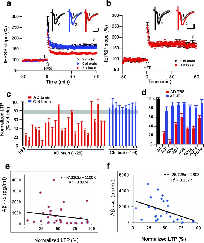
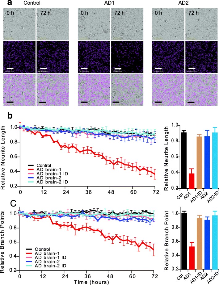
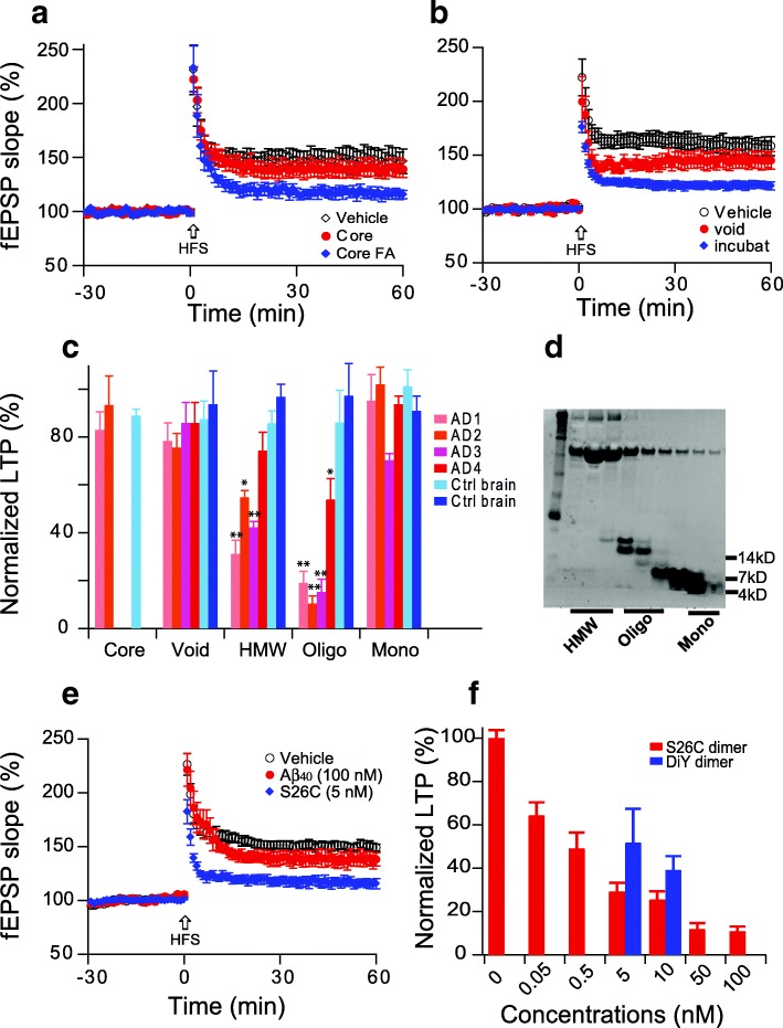
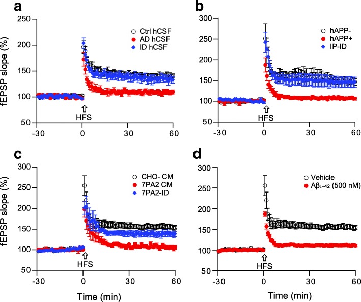
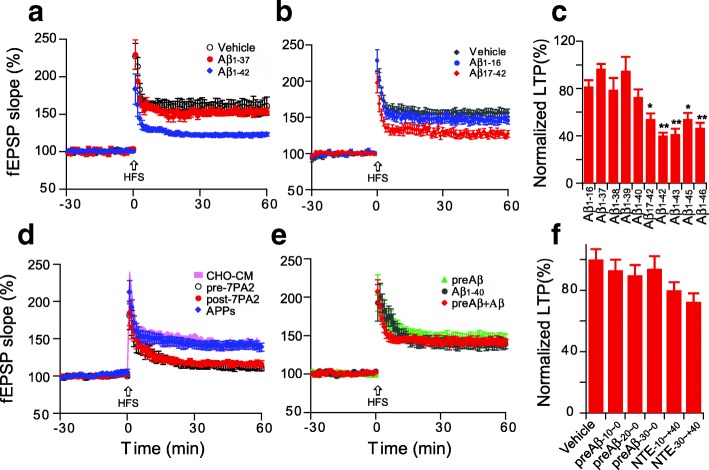
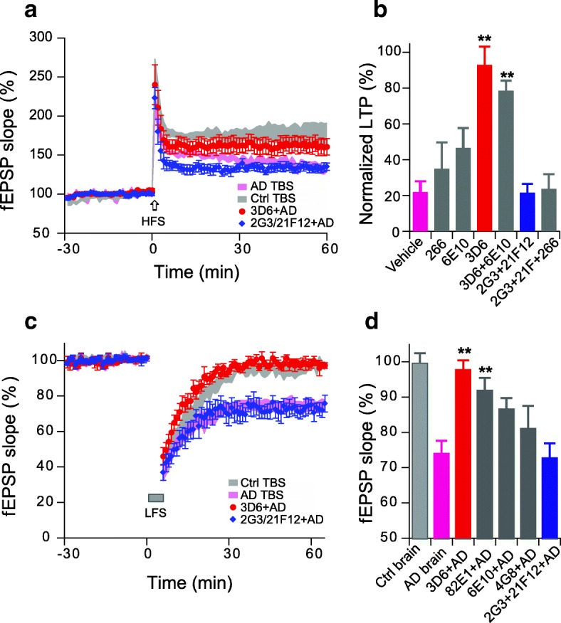
Pathologic, biochemical and genetic evidence indicates that accumulation and aggregation of amyloid β-proteins (Aβ) is a critical factor in the pathogenesis of Alzheimer's disease (AD). Several therapeutic interventions attempting to lower Aβ have failed to ameliorate cognitive decline in patients with clinical AD significantly, but most such approaches target only one or two facets of Aβ production/clearance/toxicity and do not consider the heterogeneity of human Aβ species. As synaptic dysfunction may be among the earliest deficits in AD, we used hippocampal long-term potentiation (LTP) as a sensitive indicator of the early neurotoxic effects of Aβ species. Here we confirmed prior findings that soluble Aβ oligomers, much more than fibrillar amyloid plaque cores or Aβ monomers, disrupt synaptic function. Interestingly, not all (84%) human AD brain extracts are able to inhibit LTP and the degree of LTP impairment by AD brain extracts does not correlate with Aβ levels detected by standard ELISAs. Bioactive AD brain extracts also induce neurotoxicity in iPSC-derived human neurons. Shorter forms of Aβ (including Aβ1-37, Aβ1-38, Aβ1-39), pre-Aβ APP fragments (- 30 to - 1) and N-terminally extended Aβs (- 30 to + 40) each showed much less synaptotoxicity than longer Aβs (Aβ1-42 - Aβ1-46). We found that antibodies which target the N-terminus, not the C-terminus, efficiently rescued Aβ oligomer-impaired LTP and oligomer-facilitated LTD. Our data suggest that preventing soluble Aβ oligomer formation and targeting their N-terminal residues with antibodies could be an attractive combined therapeutic approach.

Keywords: Alzheimer’s disease; Amyloid-beta protein; Long-term potentiation; Oligomers; Synaptic plasticity.


Conflict of interest statement

Competing interests

DJS is a director of and consultant to Prothena Biosciences. The other authors declare that they have no conflicts of interest.

Publisher’s Note

Springer Nature remains neutral with regard to jurisdictional claims in published maps and institutional affiliations.

Figures

Fig. 1
Fig. 1
Soluble Aβ extracted by homogenization in TBS from Alzheimer’s disease brain alters hippocampal long-term potentiation. (a) LTP induction after treatment with vehicle (black open circles), control brain TBS extract (blue circles) and AD brain TBS extract (red diamonds) from one AD patient. (b) LTP after treatment with TBS extracts made from another control brain (black diamonds) or another AD brain (red circles). (c) Summary data of LTP results with one representative run with plain TBS buffer (black bar), 25 AD brain TBS extracts (red) and 9 control (non-AD) brain TBS extracts (blue). All LTP results represent values at 60 min post-HFS normalized to vehicle alone at that time point. Gray horizontal bar indicates the lowest LTP level from control brain. (d) LTP summary data of the AD TBS extracts (red) and their respective immunodepleted extracts (blue). (e,f) Correlations between LTP levels at 60 min and the respective [Aβx-42] (e) and [Aβx-40] (f) levels in the AD TBS extracts
Fig. 2
Fig. 2
Soluble Aβ extracts that either do or do not block LTP affect human neurites accordingly. IncuCyte live-cell video microscopy monitored the effect of AD brain extracts on iPSC-derived neurogenin-induced human neurons (iNs). On post-induction day 21, iNs were treated with Control TBS (Control: black) or AD extracts (colors) and the neurons imaged for 72 h. (a) Phase contrast images (top panel) at 0 and 72 h were analyzed using the incuCyte NeuronTrack algorithm to identify neurites (pseudocolored pink in middle panels). Identified neurites were superimposed on the phase contrast image (bottom panel). Scale bar, 100 μm. Each well of iNs was imaged for 6 h prior to addition of the extract and NeuroTrack measured neurite length and branch points at this baseline used to normalize neurite length measured at each interval after addition of extract. (b,c) Time course of change in neurite length (b) and branch points (c) after addition of AD brain extracts (red and blue) or immunodepleted sample (light brown and light blue, respectively) when compare to control brain extract (black). Summary results at 72 h treatments are shown on right
Fig. 3
Fig. 3
Soluble Aβ oligomers inhibit hippocampal LTP. (a) Insoluble amyloid plaque cores from AD cortex fail to inhibit the LTP (red), while LTP was inhibited if an equivalent aliquot of the cores was solubilized in 88% formic acid and neutralized with NaOH (blue). (b) The void volume fraction of a Superdex 75 SEC chromatography of an AD cortex TBS extract (red) showed no significant LTP inhibition, but incubating the void volume fraction at 37 °C for 2 days released lower MW soluble Aβ oligomers that significantly impaired LTP (blue). (c) Summary LTP data of the indicated AD brain fractions (n = 6~ 8). (d) Representative western blot shows different MW SEC fractions of soluble Aβ-rich AD cortical extracts; (e) Oxidized synthetic [Aβ40-S26C]2 dimers (blue diamonds) cause significant LTP inhibition but monomeric Aβ1–40 does not (red); (f) Dose-dependent LTP inhibition by oxidized synthetic [Aβ40-S26C]2 dimers (red), and dityrosine cross-linked Aβ1–40 dimers (DiY) (blue). *: p < 0.05; **: p < 0.01
Fig. 4
Fig. 4
Soluble Aβ oligomers from other sources also inhibit hippocampal LTP. Several sources of soluble Aβ included (a) AD patient CSF; (b) APP tg mouse of AD (J20 mice); (c) cell secreted human soluble Aβ; and (d) synthetic Aβ1–42 peptide, effect on hippocampal LTP. All these impaired hippocampal LTP (red), while the inhibition of LTP by the 3 biological sources was prevented by removing soluble Aβ via immunodepletion (blue)
Fig. 5
Fig. 5
Soluble Aβ peptides with longer C-termini confer greater synaptic toxicity. (a) The short Aβ1–37 synthetic peptide did not impair hippocampal LTP at concentrations of 200 nM (red, n = 7, p > 0.05), while the same dose of the longer Aβ1–42 peptide showed significant inhibition (blue, n = 6, p < 0.001); (b) N-terminally truncated synthetic Aβ1–16 and Aβ17–42 effect on the hippocampal LTP. (c) Summary data of LTP effects of Aβ peptides of increasing lengths at 200 nM concentrations; (d) The whole 7PA2 CM as well as immunoprecipitated NTE-Aβs (black open circles, n = 7, p < 0.001) and the CM remaining CM after depletion of APPs by DE23 resin (red circles, n = 7, p < 0.001) all inhibit LTP, while the isolated APPs alone (blue diamonds, n = 7, p > 0.05) does not; (e) Treatment of slices with synthetic pre-Aβ (− 30 to − 1) does not facilitate synthetic Aβ1–40 to induce synaptotoxicity, that is to say, a synthetic APP-34 to − 1 fragment added to an Aβ1–40 peptide does not inhibit LTP (n = 6, p > 0.05); (f) Summary data of synthetic peptides containing or not various lengths (− 10. -20, − 30) of APP prior to the Aβ1–40 Asp1 start site (called “preAβ”) and N-terminal extension on Aβ1–40 do not inhibit LTP. (n = 6~ 8). *: p < 0.05; **: p < 0.01
Fig. 6
Fig. 6
N-terminal antibodies prevent interruption of hippocampal synaptic plasticity by soluble Aβ-rich brain extract. (a) Asp1 N-terminus specific Aβ antibody, 3D6 (red circles), not the C-terminal-specific antibodies 2G3 + 21F12 (blue diamonds) prevent the AD brain TBS extract (purple traces) from inhibiting LTP. (b) Summary data of antibodies to different Aβ epitopes as to their effect on AD brain TBS extract impairment of LTP (N > 6 recordings for each antibody). (c) N-terminal antibody 3D6 (red circles), not the C-terminal target antibodies, 2G3 + 21F12 (blue diamond) prevent the AD brain extract (purple traces) from facilitating LTD. (d). Summary data of antibodies to different Aβ epitopes as to their effect on the AD brain extract’s facilitation of LTD (N > 5 recordings for each antibody). *: p < 0.05; **: p < 0.01

References

    1. Ahmed M, Davis J, Aucoin D, Sato T, Ahuja S, Aimoto S, et al. Structural conversion of neurotoxic amyloid-beta(1-42) oligomers to fibrils. Nat Struct Mol Biol. 2010;17:561–567. doi: 10.1038/nsmb.1799. - DOI - PMC - PubMed
    1. Arendt T. Synaptic degeneration in Alzheimer's disease. Acta Neuropathol. 2009;118:167–179. doi: 10.1007/s00401-009-0536-x. - DOI - PubMed
    1. Bard F, Barbour R, Cannon C, Carretto R, Fox M, Games D, et al. Epitope and isotype specificities of antibodies to beta -amyloid peptide for protection against Alzheimer's disease-like neuropathology. Proc Natl Acad Sci U S A. 2003;100:2023–2028. doi: 10.1073/pnas.0436286100. - DOI - PMC - PubMed
    1. Bastrikova N, Gardner GA, Reece JM, Jeromin A, Dudek SM. Synapse elimination accompanies functional plasticity in hippocampal neurons. Proc Natl Acad Sci U S A. 2008;105:3123–3127. doi: 10.1073/pnas.0800027105. - DOI - PMC - PubMed
    1. Bateman RJ, Xiong C, Benzinger TL, Fagan AM, Goate A, Fox NC, et al. Clinical and biomarker changes in dominantly inherited Alzheimer's disease. N Engl J Med. 2012;367:795–804. doi: 10.1056/NEJMoa1202753. - DOI - PMC - PubMed

Publication types

MeSH terms