. 2018 Nov 8;15(1):310.
doi: 10.1186/s12974-018-1345-8.

Omega-3 polyunsaturated fatty acid attenuates traumatic brain injury-induced neuronal apoptosis by inducing autophagy through the upregulation of SIRT1-mediated deacetylation of Beclin-1

Affiliations

Omega-3 polyunsaturated fatty acid attenuates traumatic brain injury-induced neuronal apoptosis by inducing autophagy through the upregulation of SIRT1-mediated deacetylation of Beclin-1

Xiangrong Chen et al. J Neuroinflammation. .

Abstract

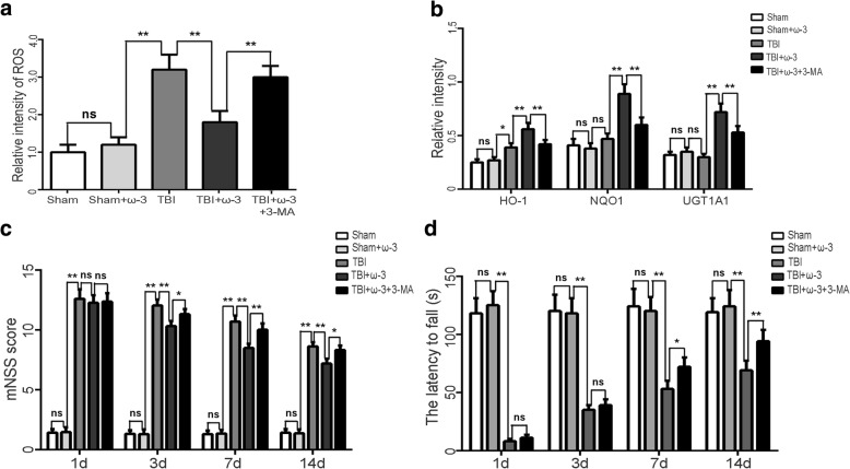
Background: Enhancing autophagy after traumatic brain injury (TBI) may decrease the expression of neuronal apoptosis-related molecules. Autophagy-mediated neuronal survival is regulated by the sirtuin family of proteins (SIRT). Omega-3 polyunsaturated fatty acids (ω-3 PUFA) are known to have antioxidative and anti-inflammatory effects. We previously demonstrated that ω-3 PUFA supplementation attenuated neuronal apoptosis by modulating the neuroinflammatory response through SIRT1-mediated deacetylation of the HMGB1/NF-κB pathway, leading to neuroprotective effects following experimental traumatic brain injury (TBI). However, no studies have elucidated if the neuroprotective effects of ω-3 PUFAs against TBI-induced neuronal apoptosis are modulated by SIRT1-mediated deacetylation of the autophagy pathway.

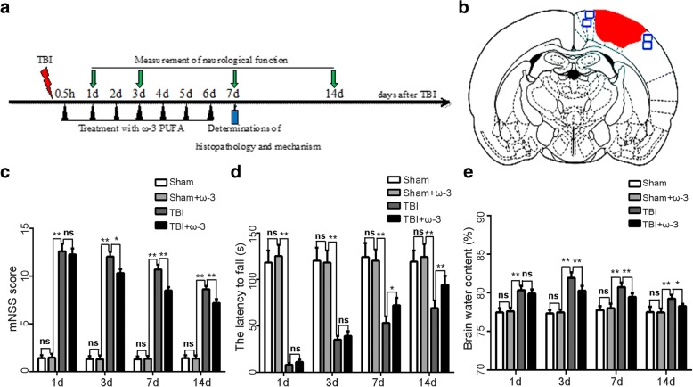
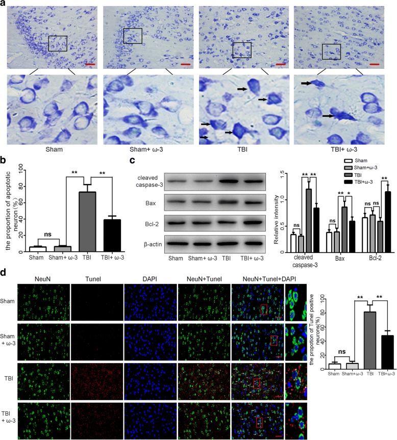
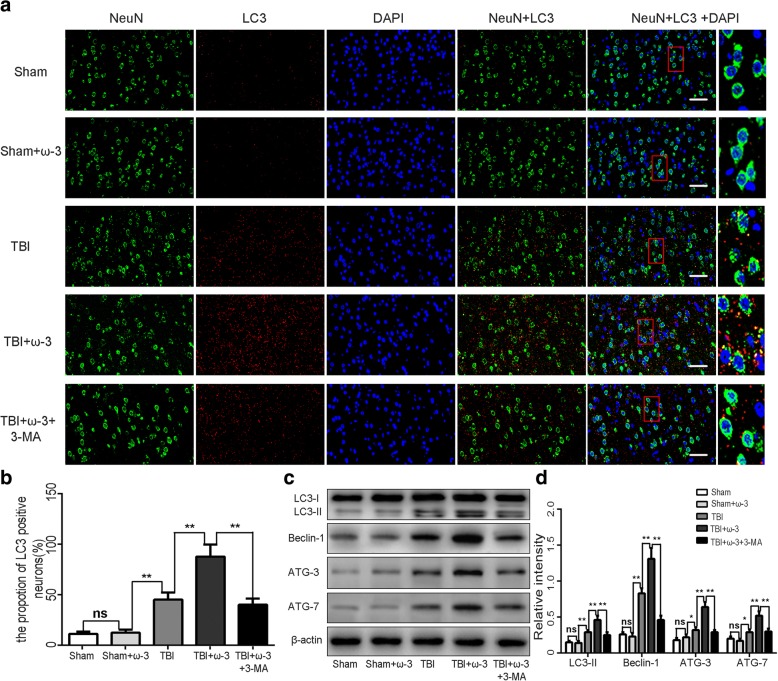
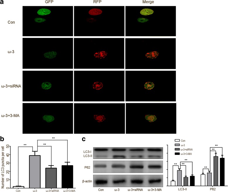
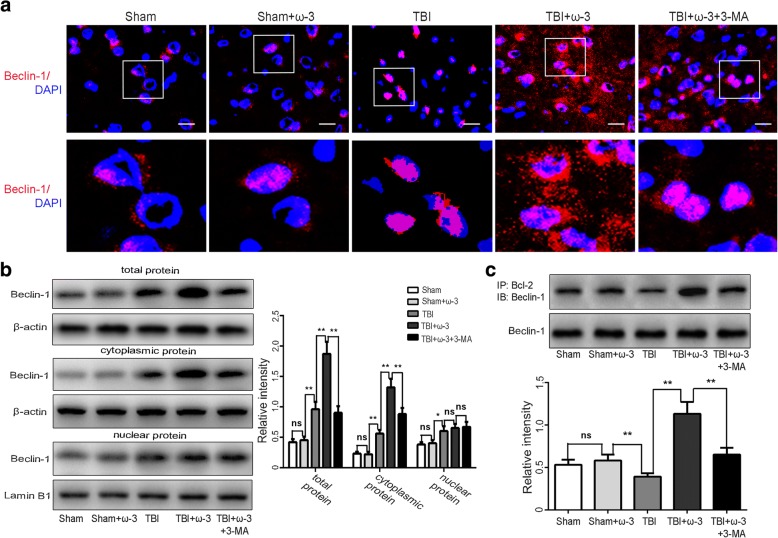
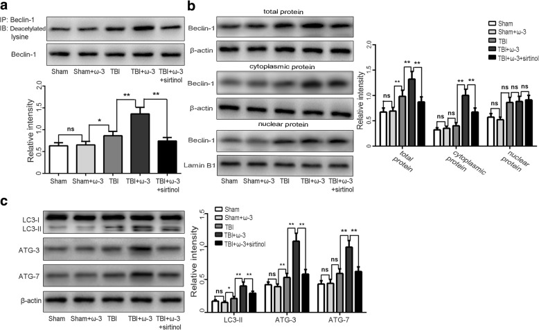
Methods: The Feeney DM TBI model was adopted to induce TBI rats. Modified neurological severity scores, the rotarod test, brain water content, and Nissl staining were employed to determine the neuroprotective effects of ω-3 PUFA supplementation. Immunofluorescent staining and western blot analysis were used to detect Beclin-1 nuclear translocation and autophagy pathway activation. The impact of SIRT1 deacetylase activity on Beclin-1 acetylation and the interaction between cytoplasmic Beclin-1 and Bcl-2 were assessed to evaluate the neuroprotective effects of ω-3 PUFAs and to determine if these effects were dependent on SIRT1-mediated deacetylation of the autophagy pathway in order to gain further insight into the mechanisms underlying the development of neuroprotection after TBI.

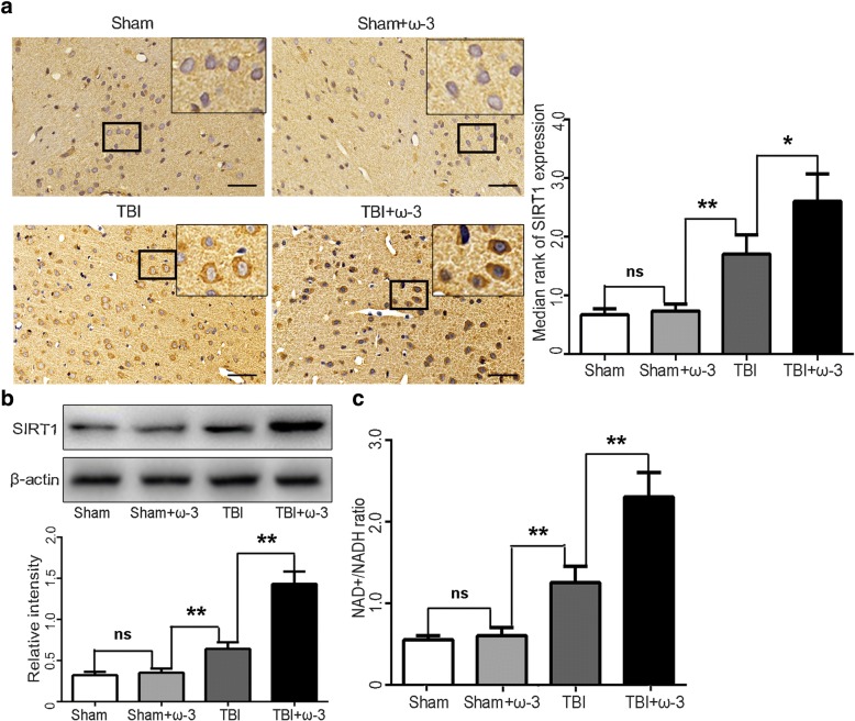
Results: ω-3 PUFA supplementation protected neurons against TBI-induced neuronal apoptosis via enhancement of the autophagy pathway. We also found that treatment with ω-3 PUFA significantly increased the NAD+/NADH ratio and SIRT1 activity following TBI. In addition, ω-3 PUFA supplementation increased Beclin-1 deacetylation and its nuclear export and induced direct interactions between cytoplasmic Beclin-1 and Bcl-2 by increasing SIRT1 activity following TBI. These events led to the inhibition of neuronal apoptosis and to neuroprotective effects through enhancing autophagy after TBI, possibly due to elevated SIRT1.

Conclusions: ω-3 PUFA supplementation attenuated TBI-induced neuronal apoptosis by inducing the autophagy pathway through the upregulation of SIRT1-mediated deacetylation of Beclin-1.

Keywords: Apoptosis; Autophagy; Omega-3 polyunsaturated fatty acid; Traumatic brain injury.


Conflict of interest statement

Ethics approval and consent to participate

The experimental protocols in the present study including all surgical procedures and animal usages conformed to the guidelines for the care and use of laboratory animals by the National Institutes of Health (NIH) and were approved by the Fujian Medical University Experimental Animal Ethics Committee (Fuzhou, China).

Consent for publication

Consent for publication is not applicable for this manuscript.

Competing interests

The authors declare that they have no competing interests.

Publisher’s Note

Springer Nature remains neutral with regard to jurisdictional claims in published maps and institutional affiliations.

Figures

Fig. 1
Fig. 1
ω-3 PUFA supplementation improves neurological function and reduces brain edema after TBI. a Experimental scheme of ω-3 PUFA supplementation after TBI. b A schematic of a brain section after TBI. Areas in red refer to lesioned sites and areas in blue refer to sample points. c ω-3 PUFA supplementation improved neurological functions 3 days after TBI (10.31 ± 0.43 vs 12.03 ± 0.53, p < 0.05). d Rats in the TBI+ω-3 PUFA group showed significantly improved rotarod performances than rats in the TBI groups from day 7 after TBI (72.01 ± 8.21 vs 53.11 ± 7.13, p < 0.05). e ω-3 PUFA supplementation decreased brain water content 3 days after TBI (80.26% ± 0.61% vs 81.92% ± 0.72%, p < 0.05). Values are expressed as mean ± standard deviation (n = 6 per group). N.S., p > 0.05, *p < 0.05, **p < 0.01
Fig. 2
Fig. 2
ω-3 PUFA supplementation protects neurons against TBI-induced neuronal apoptosis in the lesioned cortex 7 day after TBI. a, b The sham group and the sham+ω-3 PUFA group had very low fractions of apoptotic neurons. The percentage of apoptotic cells was higher in the TBI group than in the sham group (p < 0.05); the apoptotic fraction was significantly lower in the TBI+ω-3 PUFA group than in the TBI group (39.19% ± 4.72% vs 73.42% ± 9.36%, p < 0.05). Representative photomicrographs of Nissl-stained neurons are shown; arrows indicate apoptotic neurons. c Western blot analyses revealed that TBI resulted in the upregulation of apoptotic factors in the cortex; however, compared with the TBI group, cleaved caspase-3 and Bax levels were decreased, whereas the anti-apoptotic factor, Bcl-2, was increased in TBI+ω-3 PUFA group (p < 0.05). d TUNEL staining demonstrated that TUNEL-positive neurons were significantly decreased in the TBI+ω-3 group compared with the TBI group (47.72% ± 6.90% vs 81.41% ± 9.78%, p < 0.05). Representative photomicrographs of TUNEL-positive neurons are shown (× 400); arrows indicate apoptotic neurons. Values are expressed as mean ± standard deviation (n = 6 per group). N.S., p > 0.05, *p < 0.05, **p < 0.01. Scale bars = 50 μm
Fig. 3
Fig. 3
(a,b) Immunofluorescence staining showed that LC-positive neurons significantly increased following ω-3 PUFA supplementation. While, the autophagic inhibitor, 3-MA, attenuated LC-positive neuron expressions. Representative photomicrographs of LC-positive neurons are shown (× 400). (c,d) Western blot analysis showed that expression levels of autophagic markers (LC3-II, Beclin-1, ATG-3, and ATG-7) were dramatically increased in the TBI+ω-3 PUFA group 7 day after TBI (p < 0.05), while 3-MA inhibited ω-3 PUFA-induced autophagy responses. Values are expressed as mean ± standard deviation (n = 6 per group). N.S., p > 0.05, *p < 0.05, **p < 0.01. Scale bars = 50 μm
Fig. 4
Fig. 4
ω-3 PUFA supplementation promotes autophagic flux on rat hippocampal neurons in vitro. a Autophagy markers LC3 with GFP and RFP protein which indicate real-time autophagy flux levels were imaged by confocal microscope (× 400). Representative photomicrographs of autophagy flux in neurons are shown. b Bar graphs displayed the mean ± standard deviation of the LC3 puncta per cell, which indicated that the values of the ω-3 related groups were significantly different from those of the control group. The control group maintained basal levels of autophagy. The ω-3 PUFA group showed a suppression of autophagy at late stages compared to the early stage suppression of the control group, while SIRT1 siRNA or autophagy inhibitor reversed ω-3 PUFA-mediated increases in autophagy. c Western blot showed a significant increased LC3 expression and decreased p62 levels in the ω-3 PUFA treatment group. However, the SIRT1 siRNA or autophagy inhibitor groups showed an increase in p62 levels but no increases in LC3 expression. Values are expressed as mean ± standard deviation (n = 6 per group). N.S., p > 0.05, *p < 0.05, **p < 0.01
Fig. 5
Fig. 5
ω-3 PUFA supplementation protects neurons via inhibition of oxidative stress in lesioned cortices. a, b ω-3 PUFA supplementation decreased ROS activity, while it significantly increased the levels the antioxidants HO-1, NQO1, and UGT1A1 in lesioned cortices. 3-MA attenuated ω-3 PUFA-induced activation of these antioxidant factors. c, d 3-MA reversed ω-3 PUFA-mediated inhibition of neuronal apoptosis and attenuated the neuroprotective effects associated with ω-3 PUFA treatment (p < 0.05). Values are expressed as mean ± standard deviation (n = 6 per group). N.S., p > 0.05, *p < 0.05, **p < 0.01
Fig. 6
Fig. 6
ω-3 PUFA supplementation enhances autophagy via promoting nuclear export of Beclin-1 in lesioned cortices 7 days after TBI. a, b Immunofluorescence staining and western blot analyses demonstrated that expression levels of Beclin-1 in the cytosol, nuclei, and in total protein from lesioned cortices increased 7 days after TBI and that ω-3 PUFA supplementation effectively increased Beclin-1 expression in the cytosol and in total protein of cells from lesioned cortices (p < 0.05), but not in nuclear protein (p > 0.05). Moreover, less cytoplasmic redistribution of nuclear Beclin-1 was found in the presence of an autophagy inhibitor, 3-MA, after TBI. Representative photomicrographs of Beclin-1-positive neurons are shown (× 400). c Co-IP assays confirmed that ω-3 PUFA supplementation significantly increased interactions between cytoplasmic Beclin-1 and Bcl-2 after the TBI, while 3-MA treatment reversed these increases. Values are expressed as mean ± standard deviation (n = 6 per group). N.S., p > 0.05, *p < 0.05, **p < 0.01. Scale bars = 50 μm
Fig. 7
Fig. 7
ω-3 PUFA supplementation elevates SIRT1 expression and deacetylase activity in lesioned cortices 7 days after TBI. a SIRT1 immunoreactivity in both neurons and microglia from lesioned cortices was significantly increased by ω-3 PUFA supplementation (2.64 ± 0.47 vs 1.74 ± 0.33, p < 0.05). b SIRT1 levels were also upregulated after ω-3 PUFA supplementation (p < 0.05). c The NAD+/NADH ratio was measured to detect SIRT1 activity. Treatment with ω-3 PUFA significantly increased the NAD+/NADH ratio (p < 0.05). Values are expressed as mean ± standard deviation (n = 6 per group). N.S., p > 0.05, *p < 0.05, **p < 0.01. Scale bars = 50 μm
Fig. 8
Fig. 8
ω-3 PUFA supplementation increases Beclin-1 deacetylation by elevating SIRT1 activity 7 days after TBI. a IP analysis showed an elevation of Beclin-1 deacetylation following ω-3 PUFA supplementation compared with the TBI group (p < 0.05). b The nuclear export of Beclin-1 induced by ω-3 PUFA supplementation was reversed by pharmacological inhibition of SIRT (p < 0.05). c Autophagy activation induced by ω-3 PUFA supplementation was reversed by pharmacological inhibition of SIRT1 (p < 0.05). Values are expressed as mean ± standard deviation (n = 6 per group). N.S., p > 0.05, *p < 0.05, **p < 0.01

References

    1. Zhang R, Liu Y, Yan K, Chen L, Chen XR, Li P, Chen FF, Jiang XD. Anti-inflammatory and immunomodulatory mechanisms of mesenchymal stem cell transplantation in experimental traumatic brain injury. J Neuroinflammation. 2013;10:106. - PMC - PubMed
    1. Hopp S, Nolte MW, Stetter C, Kleinschnitz C, Siren AL, Albert-Weissenberger C. Alleviation of secondary brain injury, posttraumatic inflammation, and brain edema formation by inhibition of factor XIIa. J Neuroinflammation. 2017;14:39. doi: 10.1186/s12974-017-0815-8. - DOI - PMC - PubMed
    1. Sinha SP, Avcu P, Spiegler KM, Komaravolu S, Kim K, Cominski T, Servatius RJ, Pang KC. Startle suppression after mild traumatic brain injury is associated with an increase in pro-inflammatory cytokines, reactive gliosis and neuronal loss in the caudal pontine reticular nucleus. Brain Behav Immun. 2017;61:353–364. doi: 10.1016/j.bbi.2017.01.006. - DOI - PubMed
    1. McKee CA, Lukens JR. Emerging roles for the immune system in traumatic brain injury. Front Immunol. 2016;7:556. doi: 10.3389/fimmu.2016.00556. - DOI - PMC - PubMed
    1. Ding K, Xu J, Wang H, Zhang L, Wu Y, Li T. Melatonin protects the brain from apoptosis by enhancement of autophagy after traumatic brain injury in mice. Neurochem Int. 2015;91:46–54. doi: 10.1016/j.neuint.2015.10.008. - DOI - PubMed

MeSH terms

LinkOut - more resources