

Evaluation of pancreatic tumor development in KPC mice using multi-parametric MRI
- PMID: 30409175
- PMCID: PMC6225661
- DOI: 10.1186/s40644-018-0172-6
Evaluation of pancreatic tumor development in KPC mice using multi-parametric MRI
Abstract
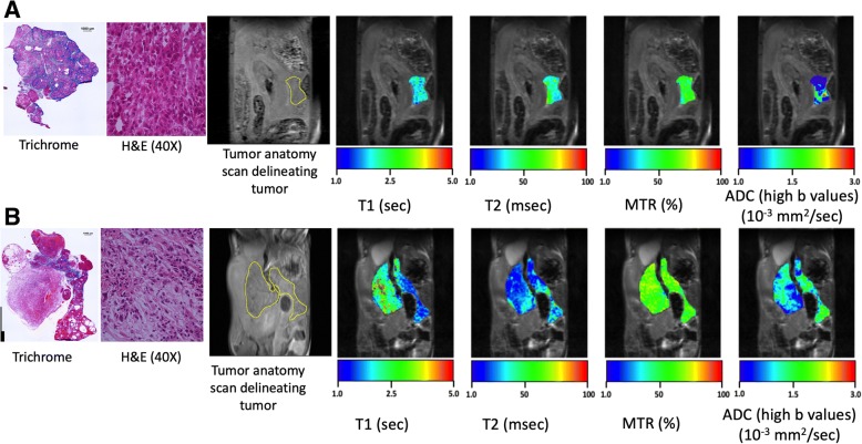
Background: Pancreatic ductal adenocarcinoma (PDA) is a fatal disease with very poor prognosis. Development of sensitive and noninvasive methods to monitor tumor progression in PDA is a critical and unmet need. Magnetic resonance imaging (MRI) can noninvasively provide information regarding underlying pathophysiological processes such as necrosis, inflammatory changes and fibrotic tissue deposition.
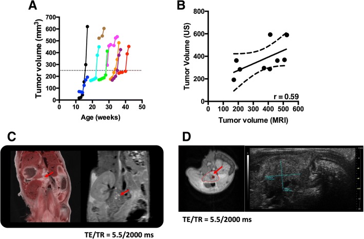
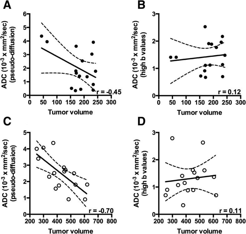
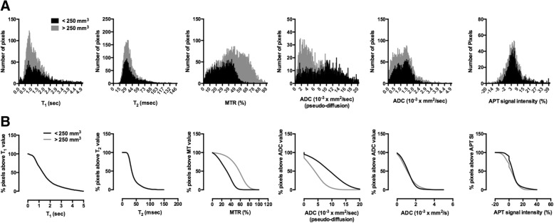
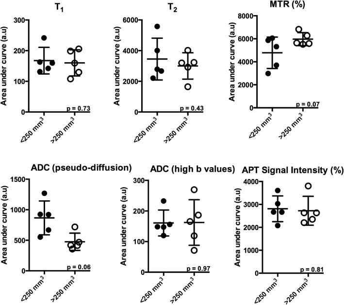
Methods: A genetically engineered KPC mouse model that recapitulates human PDA was used to characterize disease progression. MR measures of T1 and T2 relaxation times, magnetization transfer ratio (MTR), diffusion and chemical exchange saturation transfer were compared in two separate phases i.e. slow and rapid growth phase of tumor. Fibrotic tissue accumulation was assessed histologically using Masson's trichrome staining. Pearson correlation coefficient (r) was computed to assess the relationship between the fibrotic tissue accumulation and different MR parameters.
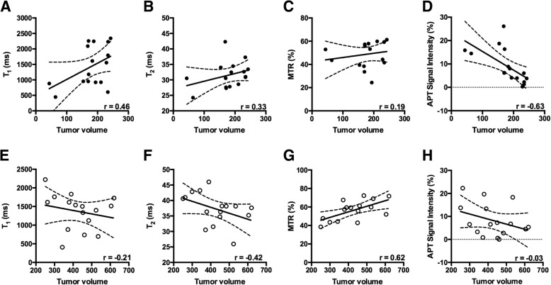
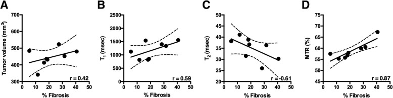
Results: There was a negative correlation between amide proton transfer signal intensity and tumor volume (r = - 0.63, p = 0.003) in the slow growth phase of the tumor development. In the terminal stage of rapid growth phase of the tumor development MTR was strongly correlated with tumor volume (r = 0.62, p = 0.008). Finally, MTR was significantly correlated with % fibrosis (r = 0.87; p < 0.01), followed by moderate correlation between tumor volume (r = 0.42); T1 (r = - 0.61), T2 (r = - 0.61) and accumulation of fibrotic tissue.
Conclusions: Here we demonstrated, using multi-parametric MRI (mp-MRI), that MRI parameters changed with tumor progression in a mouse model of PDA. Use of mp-MRI may have the potential to monitor the dynamic changes of tumor microenvironment with increase in tumor size in the transgenic KPC mouse model of pancreatic tumor.
Keywords: KPC; Multi-parametric MRI; Pancreatic ductal adenocarcinoma; Tumor microenvironment.
Conflict of interest statement
Ethics approval and consent to participate
All experiments were performed in accordance with the guidelines for the care and use of laboratory animal of the national institutes of health and with approval from our institutional animal care and use committee.
Consent for publication
Not applicable.
Competing interests
The authors declare that they have no competing interest.
Publisher’s Note
Springer Nature remains neutral with regard to jurisdictional claims in published maps and institutional affiliations.
Figures
Similar articles
-
Magnetic resonance imaging biomarkers for pulsed focused ultrasound treatment of pancreatic ductal adenocarcinoma.World J Gastroenterol. 2020 Mar 7;26(9):904-917. doi: 10.3748/wjg.v26.i9.904. World J Gastroenterol. 2020. PMID: 32206002 Free PMC article.
-
Non-Invasive Monitoring of Increased Fibrotic Tissue and Hyaluronan Deposition in the Tumor Microenvironment in the Advanced Stages of Pancreatic Ductal Adenocarcinoma.Cancers (Basel). 2022 Feb 16;14(4):999. doi: 10.3390/cancers14040999. Cancers (Basel). 2022. PMID: 35205746 Free PMC article.
-
Non-invasive dynamic monitoring initiation and growth of pancreatic tumor in the LSL-KrasG12D/+;LSL-Trp53R172H/+;Pdx-1-Cre (KPC) transgenic mouse model.J Immunol Methods. 2019 Feb;465:1-6. doi: 10.1016/j.jim.2018.11.009. Epub 2018 Nov 20. J Immunol Methods. 2019. PMID: 30468734
-
Pancreas ductal adenocarcinoma with cystic features on cross-sectional imaging: radiologic-pathologic correlation.Diagn Interv Radiol. 2018 Jan-Feb;24(1):5-11. doi: 10.5152/dir.2018.17250. Diagn Interv Radiol. 2018. PMID: 29317372 Free PMC article. Review.
-
Staging of pancreatic ductal adenocarcinoma with spiral CT and MRI.Rays. 2001 Apr-Jun;26(2):151-9. Rays. 2001. PMID: 11925786 Review.
Cited by
-
Early Differentiation of Irreversible Electroporation Ablation Regions With Radiomics Features of Conventional MRI.Acad Radiol. 2022 Sep;29(9):1378-1386. doi: 10.1016/j.acra.2021.11.020. Epub 2021 Dec 18. Acad Radiol. 2022. PMID: 34933803 Free PMC article.
-
Non-Invasive Monitoring of Stromal Biophysics with Targeted Depletion of Hyaluronan in Pancreatic Ductal Adenocarcinoma.Cancers (Basel). 2019 Jun 4;11(6):772. doi: 10.3390/cancers11060772. Cancers (Basel). 2019. PMID: 31167451 Free PMC article.
-
Multiparametric Characterization of the DSL-6A/C1 Pancreatic Cancer Model in Rats.Cancers (Basel). 2024 Apr 17;16(8):1535. doi: 10.3390/cancers16081535. Cancers (Basel). 2024. PMID: 38672617 Free PMC article.
-
Multiparametric MRI enables for differentiation of different degrees of malignancy in two murine models of breast cancer.Front Oncol. 2022 Nov 2;12:1000036. doi: 10.3389/fonc.2022.1000036. eCollection 2022. Front Oncol. 2022. PMID: 36408159 Free PMC article.
-
A Brief History and Future Prospects of CEST MRI in Clinical Non-Brain Tumor Imaging.Int J Mol Sci. 2021 Oct 26;22(21):11559. doi: 10.3390/ijms222111559. Int J Mol Sci. 2021. PMID: 34768990 Free PMC article. Review.
References
-
- Yang S, Wang X, Contino G, Liesa M, Sahin E, Ying H, Bause A, Li Y, Stommel JM, Dell'antonio G, Mautner J, Tonon G, Haigis M, Shirihai OS, Doglioni C, Bardeesy N, Kimmelman AC. Pancreatic cancers require autophagy for tumor growth. Genes Dev. 2011;25(7):717–729. doi: 10.1101/gad.2016111. - DOI - PMC - PubMed
-
- Cui JH, Kruger U, Vogel I, Luttges J, Henne-Bruns D, Kremer B, Kalthoff H. Intact tissue of gastrointestinal cancer specimen orthotopically transplanted into nude mice. Hepatogastroenterology. 1998;45(24):2087–2096. - PubMed
-
- Tan MH, Chu TM. Characterization of the tumorigenic and metastatic properties of a human pancreatic tumor cell line (AsPC-1) implanted orthotopically into nude mice. Tumour Biol. 1985;6(1):89–98. - PubMed
MeSH terms
Grants and funding
LinkOut - more resources
Full Text Sources
Medical
