

Altered functional connectivity between sub-regions in the thalamus and cortex in schizophrenia patients measured by resting state BOLD fMRI at 7T
- PMID: 30409697
- PMCID: PMC6500777
- DOI: 10.1016/j.schres.2018.10.016
Altered functional connectivity between sub-regions in the thalamus and cortex in schizophrenia patients measured by resting state BOLD fMRI at 7T
Abstract
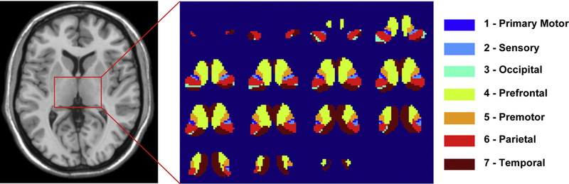
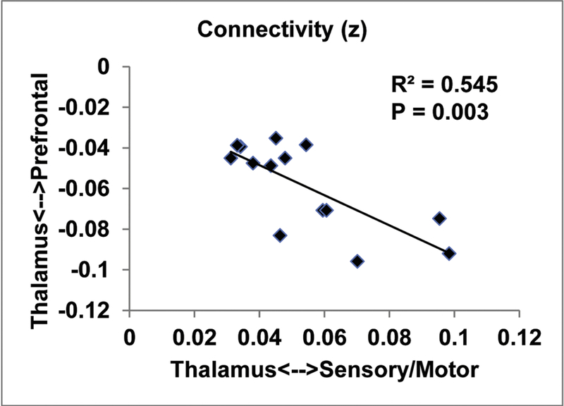
The thalamus is a small brain structure that relays neuronal signals between subcortical and cortical regions. Abnormal thalamocortical connectivity in schizophrenia has been reported in previous studies using blood-oxygenation-level-dependent (BOLD) functional MRI (fMRI) performed at 3T. However, anatomically the thalamus is not a single entity, but is subdivided into multiple distinct nuclei with different connections to various cortical regions. We sought to determine the potential benefit of using the enhanced sensitivity of BOLD fMRI at ultra-high magnetic field (7T) in exploring thalamo-cortical connectivity in schizophrenia based on subregions in the thalamus. Seeds placed in thalamic subregions of 14 patients and 14 matched controls were used to calculate whole-brain functional connectivity. Our results demonstrate impaired thalamic connectivity to the prefrontal cortex and the cerebellum, but enhanced thalamic connectivity to the motor/sensory cortex in schizophrenia. This altered functional connectivity significantly correlated with disease duration in the patients. Remarkably, comparable effect sizes observed in previous 3T studies were detected in the current 7T study with a heterogeneous and much smaller cohort, providing evidence that ultra-high field fMRI may be a powerful tool for measuring functional connectivity abnormalities in schizophrenia. Further investigation with a larger cohort is merited to validate the current findings.
Keywords: Biomarker; High field; Imaging; Psychosis; Thalamus.
Copyright © 2018 Elsevier B.V. All rights reserved.
Conflict of interest statement
Conflict of interest
Equipment used in the study was manufactured by Philips. Peter C.M. van Zijl receives grant support from Philips, is a paid lecturer for Philips, and is the inventor of technology that is licensed to Philips. This arrangement has been approved by Johns Hopkins in accordance with its conflict of interest policies.
Figures


References
-
- Anticevic A, Haut K, Murray JD, Repovs G, Yang GJ, Diehl C, McEwen SC, Bearden CE, Addington J, Goodyear B, Cadenhead KS, Mirzakhanian H, Cornblatt BA, Olvet D, Mathalon DH, McGlashan TH, Perkins DO, Belger A, Seidman LJ, Tsuang MT, van Erp TG, Walker EF, Hamann S, Woods SW, Qiu M, Cannon TD, 2015. Association of Thalamic Dysconnectivity and Conversion to Psychosis in Youth and Young Adults at Elevated Clinical Risk. JAMA psychiatry 72(9), 882–891. - PMC - PubMed
-
- Behrens TE, Johansen-Berg H, Woolrich MW, Smith SM, Wheeler-Kingshott CA, Boulby PA, Barker GJ, Sillery EL, Sheehan K, Ciccarelli O, Thompson AJ, Brady JM, Matthews PM, 2003a. Non-invasive mapping of connections between human thalamus and cortex using diffusion imaging. Nat Neurosci 6(7), 750–757. - PubMed
-
- Behrens TE, Woolrich MW, Jenkinson M, Johansen-Berg H, Nunes RG, Clare S, Matthews PM, Brady JM, Smith SM, 2003b. Characterization and propagation of uncertainty in diffusion-weighted MR imaging. Magn Reson Med 50(5), 1077–1088. - PubMed
Publication types
MeSH terms
Substances
Grants and funding
LinkOut - more resources
Full Text Sources
Medical
