. 2019 Apr;104(4):717-728.
doi: 10.3324/haematol.2018.198267. Epub 2018 Nov 8.

A new BCR-ABL1 Drosophila model as a powerful tool to elucidate the pathogenesis and progression of chronic myeloid leukemia

Affiliations

A new BCR-ABL1 Drosophila model as a powerful tool to elucidate the pathogenesis and progression of chronic myeloid leukemia

Roberto Bernardoni et al. Haematologica. 2019 Apr.

Abstract

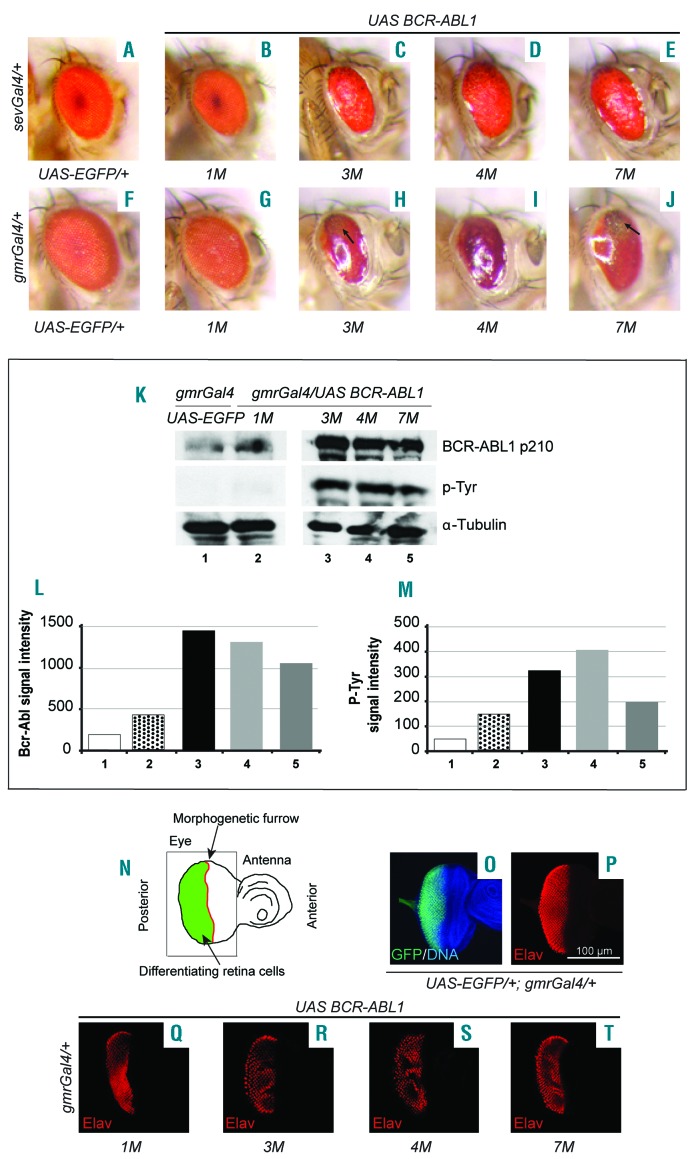
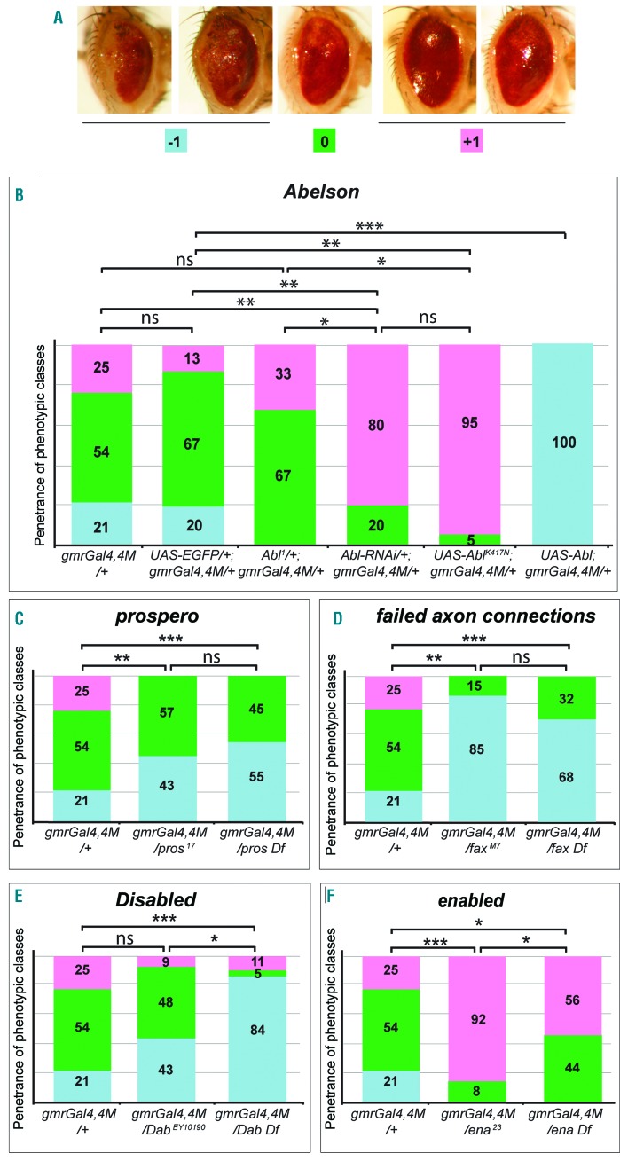
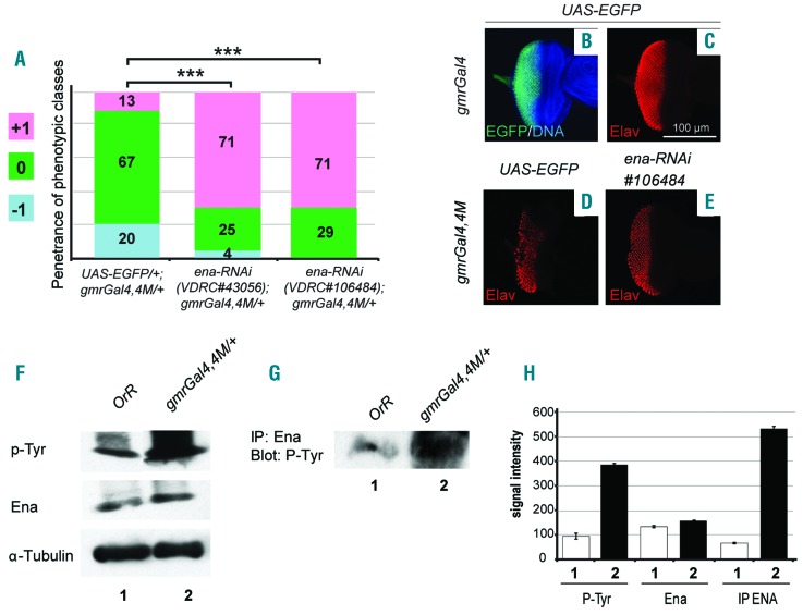
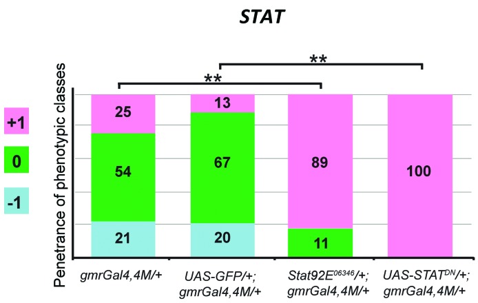
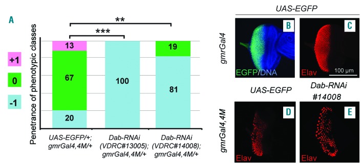
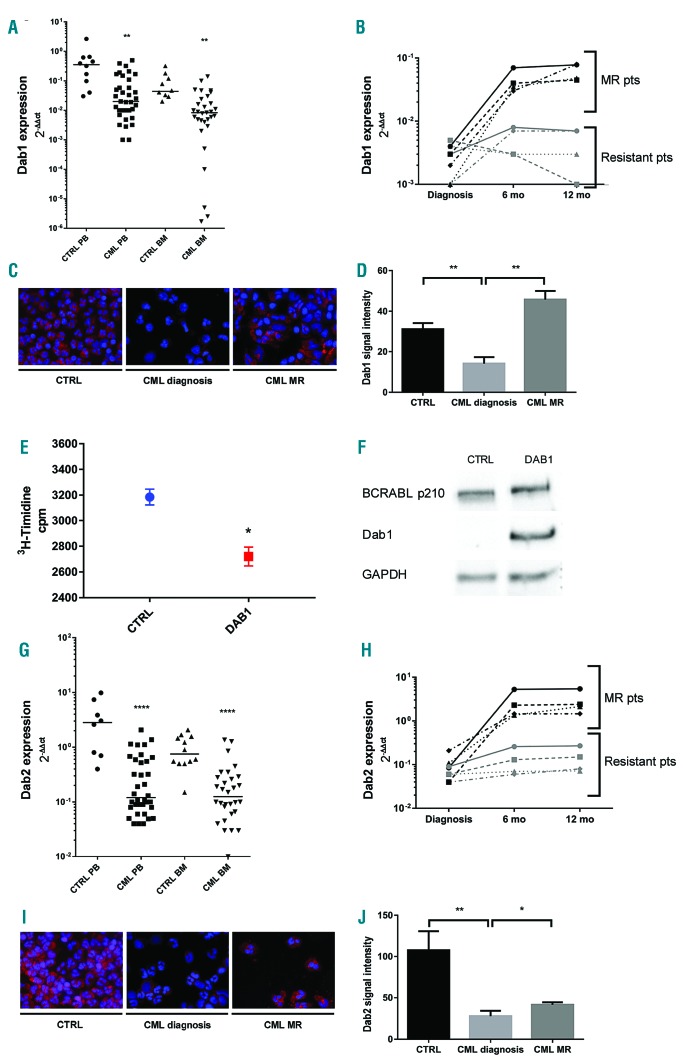
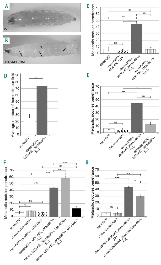
The oncoprotein BCR-ABL1 triggers chronic myeloid leukemia. It is clear that the disease relies on constitutive BCR-ABL1 kinase activity, but not all the interactors and regulators of the oncoprotein are known. We describe and validate a Drosophila leukemia model based on inducible human BCR-ABL1 expression controlled by tissue-specific promoters. The model was conceived to be a versatile tool for performing genetic screens. BCR-ABL1 expression in the developing eye interferes with ommatidia differentiation and expression in the hematopoietic precursors increases the number of circulating blood cells. We show that BCR-ABL1 interferes with the pathway of endogenous dAbl with which it shares the target protein Ena. Loss of function of ena or Dab, an upstream regulator of dAbl, respectively suppresses or enhances both the BCR-ABL1-dependent phenotypes. Importantly, in patients with leukemia decreased human Dab1 and Dab2 expression correlates with more severe disease and Dab1 expression reduces the proliferation of leukemia cells. Globally, these observations validate our Drosophila model, which promises to be an excellent system for performing unbiased genetic screens aimed at identifying new BCR-ABL1 interactors and regulators in order to better elucidate the mechanism of leukemia onset and progression.


Figures

Figure 1.
Figure 1.
BCR-ABL1 expression in the developing eye cells affects photoreceptor differentiation. (A-E) Adult eyes expressing EGFP (A) or BCR-ABL1 in four independent transgenic lines, 1M (B), 3M (C), 4M (D), or 7M (E), in a subset of differentiating photoreceptor cells under the control of the sevenlessGal4 driver construct. (C-E) High levels of BCR-ABL1 induce a “rough” eye phenotype due to impairment of cell differentiation. (F-J) Adult eyes expressing EGFP (F) or BCR-ABL1 (G-J) in all differentiating eye cells under the control of the gmrGal4 driver construct. (H-J) BCR-ABL1 expressed at high level in all differentiating eye cells profoundly disrupts ommatidia development inducing a “glazed” phenotype, depigmented area and the appearance of extra bristles (black arrows). (K-M) Quantification of BCR-ABL1 expression (K,L) and tyrosine-phosphorylation (K,M) in protein extracts from adult heads of flies expressing either EGFP (lane 1) or BCR-ABL1 in independent transgenic fly lines (lanes 2–5). The protein extracts were probed with antibodies raised against BCR, phosphorylated tyrosine residues (p-Tyr) or α–tubulin as the loading control. (N) Schematic of the eye-antenna imaginal disc from a late third instar larva; the positions of the eye and antenna primordia and of the morphogenetic furrows are indicated. The eye imaginal disc area posterior to the morphogenetic furrow, made of cells committed to terminal differentiation, is indicated in green. The thin black square indicates the region of interest shown in panels O-T. (O,P) Eye imaginal disc from wild-type late third instar larvae expressing EGFP under the control of the gmrGal4 driver in cells posterior to the morphogenetic furrow and expressing the pan-neuronal marker Elav in cells committed to terminal differentiation. (Q-T) Elav expression in eye imaginal discs from third instar larvae of the four independent transgenic lines that express BCR-ABL1 under the control of the gmrGal4 driver construct. BCR-ABL1 expression reduces the number of differentiated photoreceptors as indicated by the decrease of Elav-expressing cells.
Figure 2.
Figure 2.
BCR-ABL1 expression affects ommatidia development by altering the dAbl signaling pathway. (A) Adult eyes showing the phenotypic classes used to quantify the severity of the variable phenotype due to BCR-ABL1 expression in eye cells committed to terminal differentiation. Posterior is on the left. Class 0 (green) corresponds to the average phenotype shown by gmrGal4, UAS-BCR-ABL1 4M flies; the ommatidia are almost totally absent, the eye depigmented region is very small and the eye appears flatter than that of wild-type eyes. Class −1 (pale blue) corresponds to more severe phenotypes: the eyes are even flatter than class 0 eyes and the depigmented area is enlarged including a dorso-ventral sector in the most posterior region of the eye. Class +1 (pink) corresponds to less severe phenotypes: a few ommatidia are visible, the eyes are more bulging and the depigmented area is absent indicating better eye cell differentiation. (B-F) Adult eyes from flies of the indicated genotypes were classified to evaluate the frequency of the three phenotypic classes. (B) A piled histogram chart showing the frequencies of the different phenotypic classes in flies expressing BCR-ABL1 (gmrGal4,4M/+), co-expressing BCR-ABL1 and the EGFP (UAS-EGFP/+;gmrGal4,4M/+), expressing BCR-ABL1 but having a partial loss of the endogenous Abl gene through the heterozygous Abl1 mutation (Abl1/+;gmrGal4,4M/+), RNAi targeting Abl (Abl-RNAi/+;gmrGal4,4M/+), expression of a dominant negative dAbl mutant (UAS-AblK417N;gmrGal4,4M/+) or overexpression of the wild-type Abl protein (UAS-Abl;gmrGal4,4M/+). (C-F) Piled histogram charts showing the frequencies of the three phenotypic classes in flies expressing BCR-ABL1 (gmrGal4,4M) and heterozygous for a loss of function allele or for a deletion of genes that behave as genetic modifiers of the embryonic lethality due to Abl LOF: prospero (C), failed axon connection (D), Disabled (E) and enabled (F). The statistical comparisons were conducted using a Mann-Whitney test (*P<0.05, **P<0.01, ***P<0.001).
Figure 3.
Figure 3.
Ena loss of function suppresses the eye phenotype due to BCR-ABL1 expression which increases phosphorylation of the dAbl target Ena. (A) Piled histogram chart showing the frequencies of the three phenotypic classes in flies co-expressing BCR-ABL1 (gmrGal4,4M) and EGFP (UAS-EGFP/+;gmrGal4,4M/+), or one of two independent ena-RNAi lines (VDRC#43056 and VDRC#106484). (B,C) Eye imaginal discs from wild-type late third instar larvae expressing EGFP under the control of the gmrGal4 driver in cells posterior to the morphogenetic furrow (B) and expressing the pan-neuronal marker Elav in cells committed to terminal differentiation (C). (D,E) Elav expression in eye imaginal discs from late third instar larvae expressing BCR-ABL1 (D) or larvae co-expressing BCR-ABL1 and ena-RNAi (E) under the control of the gmrGal4 driver construct. BCR-ABL1 expression reduces the number of differentiated photoreceptors, as indicated by a decrease of Elav-expressing cells, and ena downregulation suppresses this phenotypic trait. (F-H) Quantification of Ena expression and tyrosine-phosphorylation in protein extracts (F) or Ena-immunoprecipitated proteins (G) from the heads of adult flies expressing either EGFP (lane 1) or BCR-ABL1 (lane 2). Independent loads of equal amount of protein extracts or Ena-immunoprecipitated proteins were probed with antibodies raised against phosphorylated tyrosine residues (p-Tyr), Ena or α–tubulin as loading control. (H) Average signal intensity from replica of the experiment shown in (F) and (G). Ena immunoprecipitation and probing for tyrosine-phosphorylation confirmed the increase of Ena tyrosine-phosphorylation in animals expressing BCR-ABL1. The statistical comparisons in (A) were conducted using the Mann-Whitney test (*P<0.05, **P<0.01, ***P<0.001).
Figure 4.
Figure 4.
A component of the BCR-ABL1-activated pathway in human leukemia modulates the eye phenotype in Drosophila. Piled histogram chart showing the frequencies of the three phenotypic classes in flies expressing BCR-ABL1 (gmrGal4,4M) and heterozygous for a loss of function STAT92E06346 allele or over-expressing a dominant negative allele STATDN. Reduction of the function of STAT, a gene encoding the homolog of the STAT5 protein involved in the BCR-ABL1-activated pathway in human leukemia cells, suppresses the BCR-ABL1-dependent phenotype in the fly eye. The statistical comparisons were conducted using the Mann-Whitney test (*P<0.05, **P<0.01, ***P<0.001).
Figure 5.
Figure 5.
Dab downregulation enhances the eye phenotype due to BCR-ABL1. (A) Piled histogram chart showing the frequencies of the three phenotypic classes in flies co-expressing BCR-ABL1 (gmrGal4,4M) and EGFP (UAS-EGFP/+;gmrGal4,4M/+), or one of two independent Dab-RNAi constructs (VDRC#13005, VDRC#14008). (B,C) Eye imaginal discs from wild-type late third instar larvae expressing EGFP under the control of the gmrGal4 driver in cells posterior to the morphogenetic furrow and expressing the pan-neuronal marker Elav in cells committed to terminal differentiation. (D,E) Elav expression in eye imaginal discs from late third instar larvae expressing BCR-ABL1 (D) or larvae co-expressing BCR-ABL1 and Dab-RNAi (E) under the control of the gmrGal4 driver construct. BCR-ABL1 expression reduces the number of differentiated photoreceptors, as indicated by a decrease of Elav-expressing cells, and Dab downregulation enhances this phenotypic trait. The statistical comparisons were conducted using a Mann-Whitney test (*P<0.05, **P<0.01, ***P<0.001).
Figure 6.
Figure 6.
Altered pattern of expression of the human Disabled homologs, Dab1 and Dab2, in patients with chronic myeloid leukemia. (A) Downregulation of Dab1 RNA expression in patients with chronic myeloid leukemia (CML) compared to the expression in healthy donors (CTRL). In particular we found a 1 log reduction of Dab1 expression in both peripheral blood (PB) (P<0.01) and bone marrow (BM) (P<0.01) (median values 2−ΔΔct: 0.02 versus 0.3 in PB and 0.008 versus 0.04 in BM). (B) Expression pattern of Dab1 in CML patients during molecular remission (MR) compared to that in treatment-resistant patients. (C) Immunofluorescence staining of Dab1 protein (red) in PB samples of healthy donors, CML patients at diagnosis and CML patients during MR. Nuclei are stained in blue. (D) Quantification of Dab1 protein expression in the immunofluorescence assay. (E) A 3H-thymidine proliferation assay showing a 20% reduction of cell proliferation in K562 cells transfected with Dab1 plasmid compared to control. (F) Western blot of protein extracts from K562 cells transfected with an empty vector (lane 1) and transfected with a Dab1 expression vector (lane 2), showing detectable expression of Dab1 only in K562 cells transfected with the Dab1 vector. Independent loads of equal amounts of protein extract were probed with antibodies raised against BCR, Dab1 and GAPDH as a loading control. (G) Downregulation of Dab2 RNA expression in CML patients compared to the expression in healthy donors. In particular Dab2 expression was found to be statistically decreased (P<0.0001 and P<0.0001 in PB and BM, respectively) with median values of 0.12 versus 2.8 and 0.12 versus 0.7 in PB and BM, respectively. (H) Pattern of expression of Dab2 in CML patients during MR compared to that in treatment-resistant patients. (I) Immunofluorescence staining of Dab2 protein (red) in PB samples of healthy donors, CML patients at diagnosis and CML patients during MR. Nuclei are stained in blue. (J) Quantification of Dab2 protein expression in an immunofluorescence assay. The statistical comparisons were conducted using a Student t test (*P<0.05, **P<0.01, ****P<0.0001). Bars indicate the standard error.
Figure 7.
Figure 7.
BCR-ABL1 expression in the hematopoietic precursor cells of the lymph gland impairs Drosophila blood cell homeostasis increasing the number of circulating blood cells. (A) A w1118 mid-L3 instar larva used as the wild-type control. (B) A mid-L3 larva conditionally expressing BCR-ABL1 in the hematopoietic precursors of the lymph gland medullary zone under the control of the domelessGal4 driver construct (dome:GFP/+;BCR-ABL1_3M,tub80TS/+). BCR-ABL1 expression was induced in stage L2 or early-L3 larvae by exposing the animals to 29°C during the indicated larval instars to disrupt the ability of the temperature-sensitive Gal80 mutant to inhibit Gal4 transactivation activity. The black arrows in (B) point to melanotic nodules. Anterior is on the left. (C) Penetrance of the melanotic nodule phenotype in mid-L3 control larvae expressing GFP under the control of the domelessGal4 driver (domeGFP), in larvae constitutively expressing a kinase-dead BCR-ABL1 mutant protein (dome:GFP/+;BCR-ABL1 KD/+) and in larvae in which BCR-ABL1 (dome:GFP/+;BCR-ABL1_3M,tub80TS/+) expression was induced starting from the L2 (L2) or from the early-L3 (eL3) instars. (D) Evaluation of the average number of hemocytes per field after bleeding of dome:GFP/+;BCR-ABL1_3M,tub80TS/+ and dome:GFP/+ larvae. BCR-ABL1 expression induces the appearance of melanotic nodules and this correlates with an increase of circulating hemocytes. (E) Penetrance of the melanotic nodule phenotype in mid-L3 control larvae (dome:GFP), in larvae expressing Abl-RNAi (dome/Abl-RNAi), in larvae in which BCR-ABL1 alone (dome:GFP/+;BCR-ABL1_3M,tub80TS/+) or together with Abl-RNAi (dome/Abl-RNAi;BCR-ABL1_3M,tub80TS/+) is expressed from the L2 instar. (F) Penetrance of the melanotic nodule phenotype in mid-L3 control larvae (dome:GFP), in larvae expressing Dab-RNAi (dome/+;Dab-RNAi/+), in larvae conditionally expressing the Dab protein (dome:GFP/+;tub80TS/+;UAS-Dab/+), and in larvae in which BCR-ABL1 alone (dome:GFP/+;BCR-ABL1_3M,tub80TS/+) or together with either Dab-RNAi (dome/+;BCR-ABL1_3M,tub80TS/+;Dab-RNAi/+) or UAS-Dab (dome/+;BCR-ABL1_3M,tub80TS/+;UAS-Dab/+) is expressed from the L2 instar. (G) Penetrance of the melanotic nodule phenotype in mid-L3 control larvae (dome:GFP), in larvae expressing ena-RNAi (dome/+;ena-RNAi/+), and in larvae in which BCR-ABL1 alone (dome:GFP/+;BCR-ABL1_3M,tub80TS/+) or together with ena-RNAi (dome/+;BCR-ABL1_3M,tub80TS/ena-RNAi) is expressed from the L2 instar. The average phenotype penetrance is calculated from three independent experiments, each involving 15-95 larvae. The statistical comparisons were conducted using a Student t test (*P<0.05, **P<0.01, ***P<0.001, ns=not significant). Bars indicate the standard error.

Similar articles

Cited by

References

    1. Chereda B, Melo JV. Natural course and biology of CML. Ann Hematol. 2015;94(Suppl 2):S107–121. - PubMed
    1. Quintas-Cardama A, Cortes J. Molecular biology of bcr-abl1-positive chronic myeloid leukemia. Blood. 2009;113(8):1619–1630. - PMC - PubMed
    1. Gonzalez C. Drosophila melanogaster: a model and a tool to investigate malignancy and identify new therapeutics. Nat Rev Cancer. 2013;13(3):172–183. - PubMed
    1. Bashaw GJ, Kidd T, Murray D, Pawson T, Goodman CS. Repulsive axon guidance: Abelson and Enabled play opposing roles downstream of the roundabout receptor. Cell. 2000;101(7):703–715. - PubMed
    1. Grevengoed EE, Loureiro JJ, Jesse TL, Peifer M. Abelson kinase regulates epithelial morphogenesis in Drosophila. J Cell Biol. 2001;155(7):1185–1198. - PMC - PubMed

Publication types

MeSH terms

Substances