

Brain MR Imaging Findings in Woodhouse-Sakati Syndrome
- PMID: 30409855
- PMCID: PMC7655421
- DOI: 10.3174/ajnr.A5879
Brain MR Imaging Findings in Woodhouse-Sakati Syndrome
Abstract
Background and purpose: Woodhouse-Sakati syndrome is a rare autosomal recessive disorder characterized by hypogonadism, alopecia, diabetes mellitus, and progressive extrapyramidal signs. The disease is caused by biallelic pathogenic variants in the DCAF17 gene. The purpose of this study was to describe the spectrum of brain MR imaging abnormalities in Woodhouse-Sakati syndrome.
Materials and methods: We reviewed brain MR images of 26 patients with a clinical and genetic diagnosis of Woodhouse-Sakati syndrome (12 males, 14 females; age range, 16-45 years; mean age, 26.6 years). Follow-up studies were conducted for 6 patients.
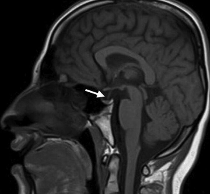
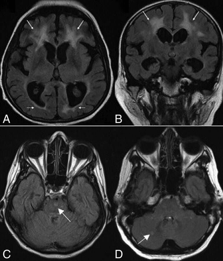
Results: All patients had abnormal MR imaging findings. The most common abnormalities were a small pituitary gland (76.9%), pronounced basal ganglia iron deposition (73%), and white matter lesions in 69.2%. White matter lesions showed frontoparietal and periventricular predominance. All white matter lesions spared subcortical U-fibers and were nonenhanced. Prominent perivascular spaces (15.3%) and restricted diffusion in the splenium of the corpus callosum (7.6%) were less frequent findings. Follow-up studies showed expansion of white matter lesions with iron deposition further involving the red nucleus and substantia nigra. Older age was associated with a more severe degree of white matter lesions (P < .001).
Conclusions: Small pituitary gland, accentuated iron deposition in the globus pallidus, and nonenhancing frontoparietal/periventricular white matter lesions were the most noted abnormalities seen in our cohort. The pattern and extent of these findings were observed to correlate with older age, reflecting a possible progressive myelin destruction and/or axonal loss. The presence of pituitary hypoplasia and white matter lesions can further distinguish Woodhouse-Sakati syndrome from other neurodegenerative diseases with brain iron accumulation subtypes.
© 2018 by American Journal of Neuroradiology.
Figures


References
-
- Bohlega SA, Alkuraya FS. Woodhouse-Sakati syndrome. 2016. August 4 In: Adam MP, Ardinger HH, Pagon RA, et al., eds. GeneReviews. https://www.ncbi.nlm.nih.gov/books/NBK378974/. Accessed May 16, 2018.
-
- Al-Semari A, Bohlega S. Autosomal-recessive syndrome with alopecia, hypogonadism, progressive extra-pyramidal disorder, white matter disease, sensory neural deafness, diabetes mellitus, and low IGF1. Am J Med Genet Part A 2007;143A:149–160 - PubMed
MeSH terms
Supplementary concepts
LinkOut - more resources
Full Text Sources
Medical