

Modulation of Mutant KrasG12D -Driven Lung Tumorigenesis In Vivo by Gain or Loss of PCDH7 Function
- PMID: 30409919
- PMCID: PMC6359939
- DOI: 10.1158/1541-7786.MCR-18-0739
Modulation of Mutant KrasG12D -Driven Lung Tumorigenesis In Vivo by Gain or Loss of PCDH7 Function
Abstract
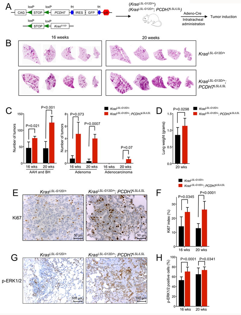
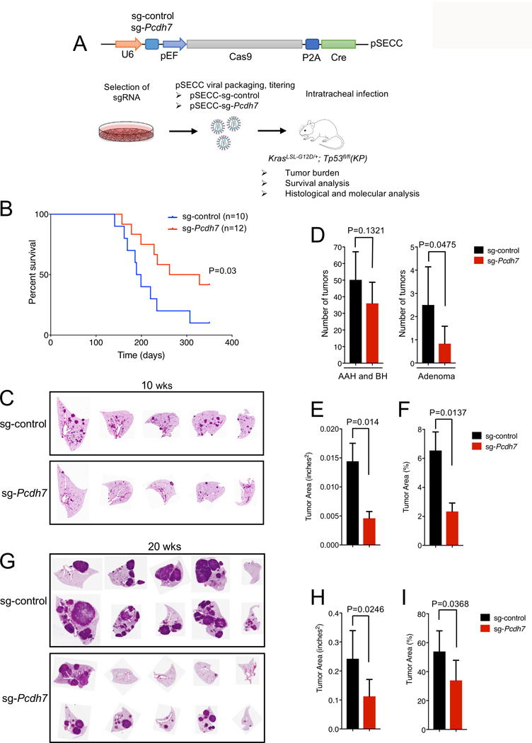
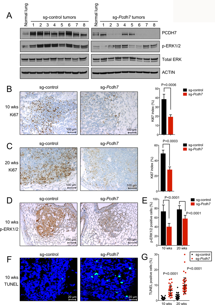
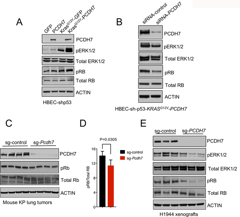
PROTOCADHERIN 7 (PCDH7), a transmembrane receptor and member of the Cadherin superfamily, is frequently overexpressed in lung adenocarcinoma and is associated with poor clinical outcome. Although PCDH7 was recently shown to promote transformation and facilitate brain metastasis in lung and breast cancers, decreased PCDH7 expression has also been documented in colorectal, gastric, and invasive bladder cancers. These data suggest context-dependent functions for PCDH7 in distinct tumor types. Given that PCDH7 is a potentially targetable molecule on the surface of cancer cells, further investigation of its role in tumorigenesis in vivo is needed to evaluate the therapeutic potential of its inhibition. Here, we report the analysis of novel PCDH7 gain- and loss-of-function mouse models and provide compelling evidence that this cell-surface protein acts as a potent lung cancer driver. Employing a Cre-inducible transgenic allele, we demonstrated that enforced PCDH7 expression significantly accelerates KrasG12D -driven lung tumorigenesis and potentiates MAPK pathway activation. Furthermore, we performed in vivo somatic genome editing with CRISPR/Cas9 in KrasLSL-G12D ; Tp53fl/fl (KP) mice to assess the consequences of PCDH7 loss of function. Inactivation of PCDH7 in KP mice significantly reduced lung tumor development, prolonged survival, and diminished phospho-activation of ERK1/2. Together, these findings establish a critical oncogenic function for PCDH7 in vivo and highlight the therapeutic potential of PCDH7 inhibition for lung cancer. Moreover, given recent reports of elevated or reduced PCDH7 in distinct tumor types, the new inducible transgenic model described here provides a robust experimental system for broadly elucidating the effects of PCDH7 overexpression in vivo. IMPLICATIONS: In this study, we establish a critical oncogenic function for PCDH7 in vivo using novel mouse models and CRISPR/Cas9 genome editing, and we validate the therapeutic potential of PCDH7 inhibition for lung cancer.
©2018 American Association for Cancer Research.
Figures
Similar articles
-
PROTOCADHERIN 7 Acts through SET and PP2A to Potentiate MAPK Signaling by EGFR and KRAS during Lung Tumorigenesis.Cancer Res. 2017 Jan 1;77(1):187-197. doi: 10.1158/0008-5472.CAN-16-1267-T. Epub 2016 Nov 7. Cancer Res. 2017. PMID: 27821484 Free PMC article.
-
Consequences of Zmat3 loss in c-MYC- and mutant KRAS-driven tumorigenesis.Cell Death Dis. 2020 Oct 20;11(10):877. doi: 10.1038/s41419-020-03066-9. Cell Death Dis. 2020. PMID: 33082333 Free PMC article.
-
PKCε Is Required for KRAS-Driven Lung Tumorigenesis.Cancer Res. 2020 Dec 1;80(23):5166-5173. doi: 10.1158/0008-5472.CAN-20-1300. Epub 2020 Sep 29. Cancer Res. 2020. PMID: 32994205 Free PMC article.
-
Regulation of EGFR signalling by palmitoylation and its role in tumorigenesis.Open Biol. 2021 Oct;11(10):210033. doi: 10.1098/rsob.210033. Epub 2021 Oct 6. Open Biol. 2021. PMID: 34610265 Free PMC article. Review.
-
Modeling K-Ras-driven lung adenocarcinoma in mice: preclinical validation of therapeutic targets.J Mol Med (Berl). 2016 Feb;94(2):121-35. doi: 10.1007/s00109-015-1360-5. Epub 2015 Nov 3. J Mol Med (Berl). 2016. PMID: 26526121 Review.
Cited by
-
Lactate receptor GPR81 drives breast cancer growth and invasiveness through regulation of ECM properties and Notch ligand DLL4.BMC Cancer. 2023 Nov 22;23(1):1136. doi: 10.1186/s12885-023-11631-6. BMC Cancer. 2023. PMID: 37993804 Free PMC article.
-
PP2A-Mediated GSK3β Dephosphorylation Is Required for Protocadherin-7-Dependent Regulation of Small GTPase RhoA in Osteoclasts.Cells. 2023 Jul 29;12(15):1967. doi: 10.3390/cells12151967. Cells. 2023. PMID: 37566044 Free PMC article.
-
A more novel and powerful prognostic gene signature of lung adenocarcinoma determined from the immune cell infiltration landscape.Front Surg. 2022 Oct 13;9:1015263. doi: 10.3389/fsurg.2022.1015263. eCollection 2022. Front Surg. 2022. PMID: 36311939 Free PMC article.
-
CRISPER/CAS System, a Novel Tool of Targeted Therapy of Drug-Resistant Lung Cancer.Adv Pharm Bull. 2022 Mar;12(2):262-273. doi: 10.34172/apb.2022.027. Epub 2021 Apr 3. Adv Pharm Bull. 2022. PMID: 35620343 Free PMC article. Review.
-
Identification of cold tumor induction-related markers in pancreatic cancer and the clinical implication of PCDH7.J Cancer Res Clin Oncol. 2025 Jan 24;151(2):45. doi: 10.1007/s00432-025-06095-z. J Cancer Res Clin Oncol. 2025. PMID: 39856454 Free PMC article.
References
-
- Kahr I, Vandepoele K, van Roy F. Delta-protocadherins in health and disease. Progress in molecular biology and translational science 2013;116:169–92. - PubMed
-
- van Roy F Beyond E-cadherin: roles of other cadherin superfamily members in cancer. Nature reviews Cancer 2014;14:121–34. - PubMed
-
- Vincent A, Omura N, Hong SM, Jaffe A, Eshleman J, Goggins M. Genome-wide analysis of promoter methylation associated with gene expression profile in pancreatic adenocarcinoma. Clinical cancer research : an official journal of the American Association for Cancer Research 2011;17:4341–54. - PMC - PubMed
-
- Yang X, Chen MW, Terry S, Vacherot F, Chopin DK, Bemis DL, et al. A human- and male-specific protocadherin that acts through the wnt signaling pathway to induce neuroendocrine transdifferentiation of prostate cancer cells. Cancer research 2005;65:5263–71. - PubMed
Publication types
MeSH terms
Substances
Grants and funding
LinkOut - more resources
Full Text Sources
Medical
Molecular Biology Databases
Research Materials
Miscellaneous
