. 2018 Nov 8;8(1):16529.
doi: 10.1038/s41598-018-33070-1.

FXR agonist obeticholic acid induces liver growth but exacerbates biliary injury in rats with obstructive cholestasis

Affiliations

FXR agonist obeticholic acid induces liver growth but exacerbates biliary injury in rats with obstructive cholestasis

Rowan F van Golen et al. Sci Rep. .

Abstract

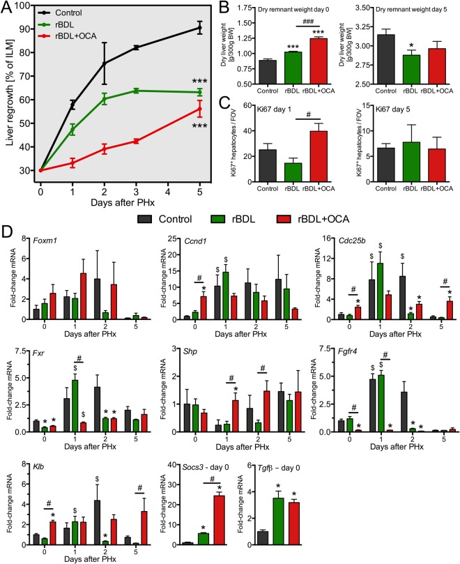
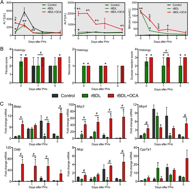
Cholestasis impairs liver regeneration following partial liver resection (PHx). Bile acid receptor farnesoid X-receptor (FXR) is a key mediator of liver regeneration. The effects of FXR agonist obeticholic acid (OCA) on liver (re)growth were therefore studied in cholestatic rats. Animals underwent sham surgery or reversible bile duct ligation (rBDL). PHx with concurrent internal biliary drainage was performed 7 days after rBDL. Animals were untreated or received OCA (10 mg/kg/day) per oral gavage from rBDL until sacrifice. After 7 days of OCA treatment, dry liver weight increased in the rBDL + OCA group, indicating OCA-mediated liver growth. Enhanced proliferation in the rBDL + OCA group prior to PHx concurred with a rise in Ki67-positive hepatocytes, elevated hepatic Ccnd1 and Cdc25b expression, and an induction of intestinal fibroblast growth factor 15 expression. Liver regrowth after PHx was initially stagnant in the rBDL + OCA group, possibly due to hepatomegaly prior to PHx. OCA increased hepatobiliary injury markers during BDL, which was accompanied by upregulation of the bile salt export pump. There were no differences in histological liver injury. In conclusion, OCA induces liver growth in cholestatic rats prior to PHx but exacerbates biliary injury during cholestasis, likely by forced pumping of bile acids into an obstructed biliary tree.


Conflict of interest statement

The authors declare no competing interests.

Figures

Figure 1
Figure 1
Obeticholic acid induces hepatocyte proliferation and hepatomegaly in cholestatic rats prior to liver resection. (A) Shows the total liver weight of controls (sham surgery) and after 7 days of rBDL with or without OCA treatment. Liver weight is expressed as percentage of body weight (A) or as dry weight per 300 g body weight (B,C) shows quantitative assessment of Ki67 positivity and (D) includes representative Ki67-stained liver sections. (E,F) Show expression of proliferation- and Fxr-related genes in liver or ileum, respectively. Levels are expressed relative to mean expression in the control rats. Abbreviations: BW = body weight; FOV = field of view; OCA = obeticholic acid; rBDL = reversible bile duct ligation (cholestasis). *Indicates p < 0.05, **indicates p < 0.01, and ***indicates p < 0.001, all versus the control group. #Indicates p < 0.05, ##indicates p < 0.01, and ###indicates p < 0.001, all between the experimental groups indicated by the solid line. N = 4–6 per group per time point.
Figure 2
Figure 2
Liver regrowth is reduced in post-cholestatic rats treated with obeticholic acid. (A) Shows liver regrowth after partial hepatectomy (PHx) in healthy rats (black line), in untreated post-cholestatic rats (rBDL, green line), and rBDL rats receiving obeticholic acid (OCA, red line). Shown in (B) are the dry weights over the remnant liver immediately (left) and five days after PHx (right). (C) Shows the number of proliferating hepatocytes on day 1 (left) and day 5 (right) after PHx. (D) Presents the hepatic expression of various proliferation and Fxr-related genes. Levels are expressed relative to mean expression in the control rats at day 0. Abbreviations: BW = body weight; rBDL = reversible bile duct ligation (cholestasis); OCA = obeticholic acid. *Indicates p < 0.05 versus the control group. #Signifies p < 0.05 between the experimental groups indicated by the solid line. $Indicates p < 0.05 versus baseline (day 0) within an experimental group. N = 4–6 per group per time point.
Figure 3
Figure 3
Obeticholic acid aggravates cholestatic injury through Bsep-mediated bile acid export. (A) Shows serum hepatobiliary injury markers, measured before (t = 0) and after partial hepatectomy (PHx) in controls rats (black line), and (post)cholestatic rats left untreated (green line) or receiving obeticholic acid (OCA, red line). (B) Shows histologic scoring of H&E-stained liver sections shown as mean score ± range. Apart from fibrosis from post-surgery day 1 onwards, no histological anomalies were observed in control rats. The scoring system and representative images are included as Supplementary Information. (C) Shows expression of genes related to hepatocyte BA homeostasis. Levels are expressed relative to mean expression in the control rats at day 0. Abbreviations: ALP = alkaline phosphatase; ALT = alanine aminotransferase; rBDL = reversible bile duct ligation (cholestasis); OCA = obeticholic acid. *Indicates p < 0.05 versus the control group and #signifies p < 0.05 versus the experimental group indicated by the solid line. N = 4–6 per group per time point.
Figure 4
Figure 4
Obeticholic acid increases biliary bile acid output in rats with obstructive cholestasis. Shown are total bile acid (BA) levels in plasma (A) and liver tissue (B), and bile collected from the extrahepatic bile duct before decompression and subsequent PHx (CD). The total BA data for the control group and the untreated BDL group were published previously. Data were obtained after seven days of BDL (i.e., prior to PHx). Abbreviations: rBDL = reversible bile duct ligation; OCA = obeticholic acid. ***Indicates p < 0.001 versus the control group. #Indicates p < 0.05 and ###indicates p < 0.001 between the experimental groups indicated by the solid line. N = 4–6 per group per time point.

Similar articles

Cited by

References

    1. Taub R. Liver regeneration: from myth to mechanism. Nat Rev Mol Cell Biol. 2004;5:836–847. doi: 10.1038/nrm1489. - DOI - PubMed
    1. de Meijer VE, Kalish BT, Puder M, Ijzermans JN. Systematic review and meta-analysis of steatosis as a risk factor in major hepatic resection. Br J Surg. 2010;97:1331–1339. doi: 10.1002/bjs.7194. - DOI - PubMed
    1. Farges O, et al. Multicentre European study of preoperative biliary drainage for hilar cholangiocarcinoma. Br J Surg. 2013;100:274–283. doi: 10.1002/bjs.8950. - DOI - PubMed
    1. Wiggers JK, et al. Postoperative Mortality after Liver Resection for Perihilar Cholangiocarcinoma: Development of a Risk Score and Importance of Biliary Drainage of the Future Liver Remnant. J Am Coll Surg. 2016;223:321–331.e1. doi: 10.1016/j.jamcollsurg.2016.03.035. - DOI - PMC - PubMed
    1. Schaap FG, Trauner M, Jansen PLM. Bile acid receptors as targets for drug development. Nature Reviews Gastroenterology & Hepatology. 2013;11:55–67. doi: 10.1038/nrgastro.2013.151. - DOI - PubMed

Publication types

MeSH terms