

Structural basis for the recognition of sulfur in phosphorothioated DNA
- PMID: 30409991
- PMCID: PMC6224610
- DOI: 10.1038/s41467-018-07093-1
Structural basis for the recognition of sulfur in phosphorothioated DNA
Abstract
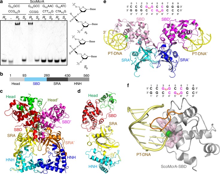
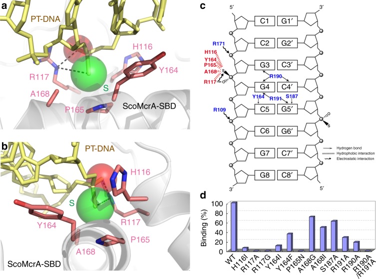
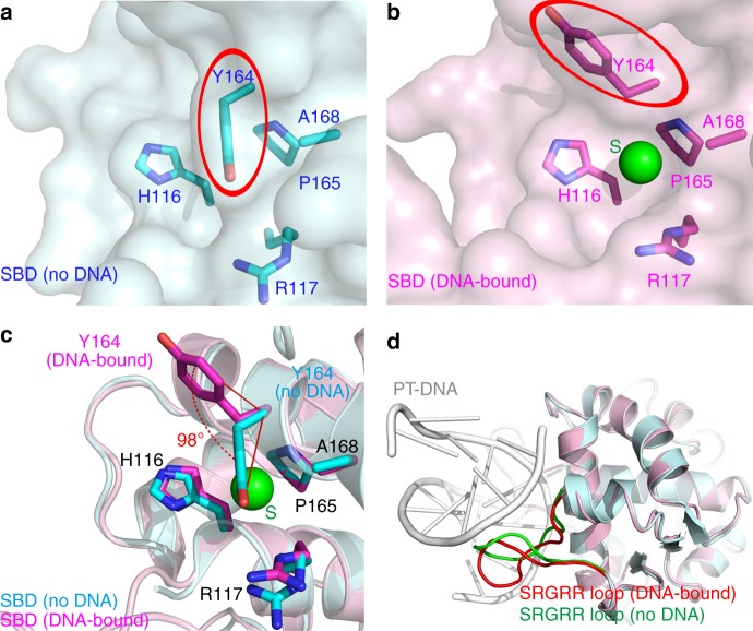
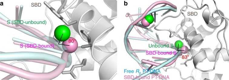
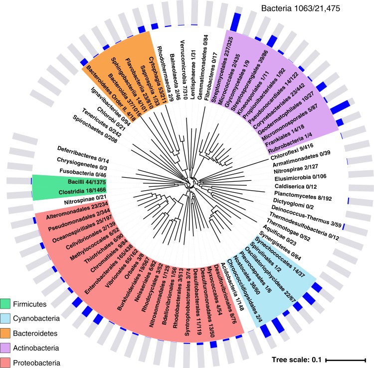
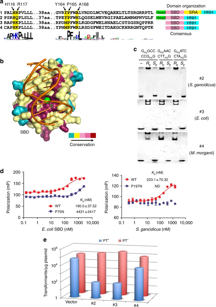
There have been very few reports on protein domains that specifically recognize sulfur. Here we present the crystal structure of the sulfur-binding domain (SBD) from the DNA phosphorothioation (PT)-dependent restriction endonuclease ScoMcrA. SBD contains a hydrophobic surface cavity that is formed by the aromatic ring of Y164, the pyrolidine ring of P165, and the non-polar side chains of four other residues that serve as lid, base, and wall of the cavity. The SBD and PT-DNA undergo conformational changes upon binding. The S187RGRR191 loop inserts into the DNA major groove to make contacts with the bases of the GPSGCC core sequence. Mutating key residues of SBD impairs PT-DNA association. More than 1000 sequenced microbial species from fourteen phyla contain SBD homologs. We show that three of these homologs bind PT-DNA in vitro and restrict PT-DNA gene transfer in vivo. These results show that SBD-like PT-DNA readers exist widely in prokaryotes.
Conflict of interest statement
The authors declare no competing interests.
Figures
Similar articles
-
DNA backbone interactions impact the sequence specificity of DNA sulfur-binding domains: revelations from structural analyses.Nucleic Acids Res. 2020 Sep 4;48(15):8755-8766. doi: 10.1093/nar/gkaa574. Nucleic Acids Res. 2020. PMID: 32621606 Free PMC article.
-
Molecular recognition between bacterial phosphorothioate DNA and sulfur-binding domain (SBD): competition between the water cage and chalcogen-hydrophobic packet.Phys Chem Chem Phys. 2022 Apr 20;24(16):9176-9187. doi: 10.1039/d2cp00291d. Phys Chem Chem Phys. 2022. PMID: 35383346
-
Protein Domain Guided Screen for Sequence Specific and Phosphorothioate-Dependent Restriction Endonucleases.Front Microbiol. 2020 Aug 18;11:1960. doi: 10.3389/fmicb.2020.01960. eCollection 2020. Front Microbiol. 2020. PMID: 33013736 Free PMC article.
-
Three-dimensional structural views of damaged-DNA recognition: T4 endonuclease V, E. coli Vsr protein, and human nucleotide excision repair factor XPA.Mutat Res. 2000 Aug 30;460(3-4):257-75. doi: 10.1016/s0921-8777(00)00031-8. Mutat Res. 2000. PMID: 10946233 Review.
-
STAND, a class of P-loop NTPases including animal and plant regulators of programmed cell death: multiple, complex domain architectures, unusual phyletic patterns, and evolution by horizontal gene transfer.J Mol Biol. 2004 Oct 8;343(1):1-28. doi: 10.1016/j.jmb.2004.08.023. J Mol Biol. 2004. PMID: 15381417 Review.
Cited by
-
Nature Chose Phosphates and Chemists Should Too: How Emerging P(V) Methods Can Augment Existing Strategies.ACS Cent Sci. 2021 Sep 22;7(9):1473-1485. doi: 10.1021/acscentsci.1c00487. Epub 2021 Sep 10. ACS Cent Sci. 2021. PMID: 34584948 Free PMC article. Review.
-
Molecular basis of the phosphorothioation-sensing antiphage defense system IscS-DndBCDE-DndI.Nucleic Acids Res. 2024 Dec 11;52(22):13594-13604. doi: 10.1093/nar/gkae1133. Nucleic Acids Res. 2024. PMID: 39611571 Free PMC article.
-
A DNA phosphorothioation pathway via adenylated intermediate modulates Tdp machinery.Nat Chem Biol. 2025 Aug;21(8):1160-1170. doi: 10.1038/s41589-024-01832-w. Epub 2025 Jan 16. Nat Chem Biol. 2025. PMID: 39820821
-
Molecular Insight into the Recognition of DNA by the DndCDE Complex in DNA Phosphorothioation.Int J Mol Sci. 2025 Jun 16;26(12):5765. doi: 10.3390/ijms26125765. Int J Mol Sci. 2025. PMID: 40565227 Free PMC article.
-
Characterization of a promiscuous DNA sulfur binding domain and application in site-directed RNA base editing.Nucleic Acids Res. 2023 Oct 27;51(19):10782-10794. doi: 10.1093/nar/gkad743. Nucleic Acids Res. 2023. PMID: 37702119 Free PMC article.
References
Publication types
MeSH terms
Substances
LinkOut - more resources
Full Text Sources
Molecular Biology Databases
