. 2018 Nov 8;8(1):16587.
doi: 10.1038/s41598-018-34795-9.

Structural Studies based on two Lysine Dioxygenases with Distinct Regioselectivity Brings Insights Into Enzyme Specificity within the Clavaminate Synthase-Like Family

Affiliations

Structural Studies based on two Lysine Dioxygenases with Distinct Regioselectivity Brings Insights Into Enzyme Specificity within the Clavaminate Synthase-Like Family

Karine Bastard et al. Sci Rep. .

Abstract

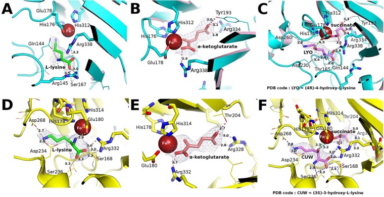
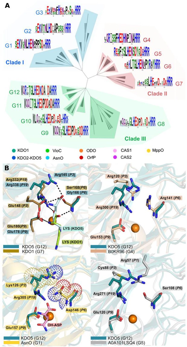
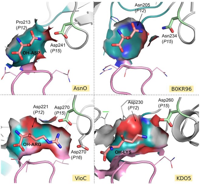
Iron(II)/α-ketoacid-dependent oxygenases (αKAOs) are enzymes that catalyze the oxidation of unactivated C-H bonds, mainly through hydroxylation. Among these, those that are active towards amino-acids and their derivatives are grouped in the Clavaminate Synthase Like (CSL) family. CSL enzymes exhibit high regio- and stereoselectivities with strict substrate specificity. This study reports the structural elucidation of two new regiodivergent members, KDO1 and KDO5, active towards lysine, and the structural and computational analysis of the whole family through modelling and classification of active sites. The structures of KDO1 and KDO5 in complex with their ligands show that one exact position in the active site controls the regioselectivity of the reaction. Our results suggest that the substrate specificity and high stereoselectivity typical of this family is linked to a lid that closes up in order to form a sub-pocket around the side chain of the substrate. This dynamic lid is found throughout the family with varying sequence and length and is associated with a conserved stable dimeric interface. Results from this study could be a starting-point for exploring the functional diversity of the CSL family and direct in vitro screening in the search for new enzymatic activities.


Conflict of interest statement

The authors declare no competing interests.

Figures

Figure 1
Figure 1
B-factor putty representation of the dimeric interface of KDO1 and KDO5. The thickness of a region is proportional to its local B-factor and thus its flexibility. (A) KDO1 dimer in shades of pink (monomers A and B of the KDO1-Fe-α-KG structure). (B) KDO5 dimer in shades of green (monomers A and B of the KDO5-Fe-α-KG structure). Helix α2, α3 and α8 at the dimeric interface, lid and adjacent loop are highlighted.
Figure 2
Figure 2
Comparison of the binding sites of KDO5 and KDO1, with the 2Fo-Fc electron density (contoured at 0.5 σ) of the bound substrates and products. (A) KDO5 in complex with L-lysine (monomer C). (B) KDO5 in complex with α-KG (monomer A). (C) KDO5 in complex with (4R)-4-hydroxy-L-lysine and succinate (monomer D). (D) KDO1 in complex with L-lysine (monomer C). (E) KDO1 in complex with α-KG (monomer B). (F) KDO1 in complex with (3S)-3-hydroxy-L-lysine and succinate (monomer B). Among the four molecules in the asymmetric unit, only the binding site with the best positioning in the electron density for the ligands has been illustrated. Identifying codes assigned for the small ligand compounds are indicated on Panels B and E.
Figure 3
Figure 3
Sequence Similarity Network (SSN) of the Clavaminate synthase-like family (IPR014503 InterPro family) from the αKAOs superfamily. Nodes represent proteins and edges are the links between those proteins that have at least 50% sequence identity. (A) Experimental data retrieved from Swissprot mapped on the SSN. KDO5 and other KDO2-4, characterized previously are colored in dark blue. (B) Annotations from Uniprot mapped on the SSN. The network is visualized with Gephi (Bastian, “Gephi: An Open Source Software for Exploring and Manipulating Networks”, AAAI Publications, Third International AAAI Conference on Weblogs and Social Media, 2009).
Figure 4
Figure 4
Dividing the CSL family into groups with similar active sites. (A) ASMC hierarchical tree containing 523 proteins, grouped in 3 clades (I, II, III). For each ASMC group (from G1 to G12), a logo sequence is depicted and represents the conservation of residues in the active site pocket. Proteins with known activities are indicated by a circle in the tree. (B) Regioselectivity among the CSL proteins. Superimposition of crystallographic structure of KDO5, chain C, in complex with lysine, with crystallographic structure of KDO1, chain C, in complex with lysine (top left), with a representant of group G4 (model with Uniprot ID B0KR96) (top right), with crystal structure of AsnO in complex with hydroxy-aspartate (PDB code 2OG7) (down left) and with a representant of G5 (model with Uniprot ID A0A101LSQ4) (down right). Residues interacting with the carboxylate and amino group of the substrates are indicated in stick, along with their position in the logo. Black dashed lines indicate interactions between substrates and proteins.
Figure 5
Figure 5
Depiction of the sub-pockets in the CSL family. For Clade I, represented by AsnO, the sub-pocket is formed by 3D motif PPSD in P12 to P15 of the active site logo. For Clade II, a representative of the G4 group, a model of protein with Uniprot ID B0KR96, was chosen. The sub-pocket is composed of residues NPIN. To get an idea of the size of substrate that can host the protein, Asn from AsnO was positioned at the center of the binding pocket. For Clade III, represented by VioC and KDO5, the sub-pocket is formed by 3D motifs DSDD and DA-D respectively. Residue in P15 of the logo is represented in green. The flexible lid is colored deep blue teal with atoms colored by elements. The flexible lid occupies a large part of the binding pocket, which is represented in surface. The adjacent loop is colored in pink.

References

    1. Sheldon RA, Woodley JM. Role of Biocatalysis in Sustainable Chemistry. Chem. Rev. 2018;118:801–838. doi: 10.1021/acs.chemrev.7b00203. - DOI - PubMed
    1. Bornscheuer UT, et al. Engineering the third wave of biocatalysis. Nature. 2012;485:185–194. doi: 10.1038/nature11117. - DOI - PubMed
    1. Wells AS, Finch GL, Michels PC, Wong JW. Use of Enzymes in the Manufacture of Active Pharmaceutical Ingredients. A Science and Safety-Based Approach To Ensure Patient Safety and Drug Quality. Org. Process Res. Dev. 2012;16:1986–1993. doi: 10.1021/op300153b. - DOI
    1. Sun H, Zhang H, Ang EL, Zhao H. Biocatalysis for the synthesis of pharmaceuticals and pharmaceutical intermediates. Bioorg. Med. Chem. 2018;16:1275–1284. doi: 10.1016/j.bmc.2017.06.043. - DOI - PubMed
    1. Wan J-P, Gan L, Liu Y. Transition metal-catalyzed C-H bond functionalization in multicomponent reactions: a tool toward molecular diversity. Org. Biomol. Chem. 2017;15:9031–9043. doi: 10.1039/C7OB02011B. - DOI - PubMed

Publication types

MeSH terms