

Improving clinical diagnosis of early-stage cutaneous melanoma based on Raman spectroscopy
- PMID: 30410059
- PMCID: PMC6265324
- DOI: 10.1038/s41416-018-0257-9
Improving clinical diagnosis of early-stage cutaneous melanoma based on Raman spectroscopy
Abstract
Background: Clinical diagnosis of early melanoma (Breslow thickness less than 0.8 mm) is crucial to disease-free survival. However, it is subjective and can be exceedingly difficult, leading to missed melanomas, or unnecessary excision of benign pigmented skin lesions. An objective technique is needed to improve the diagnosis of early melanoma.
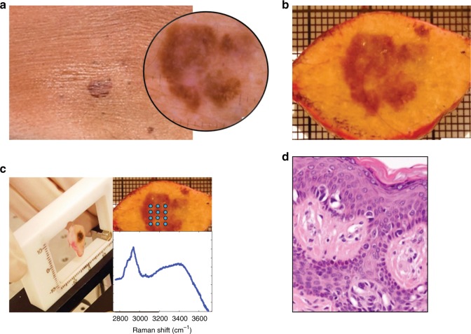
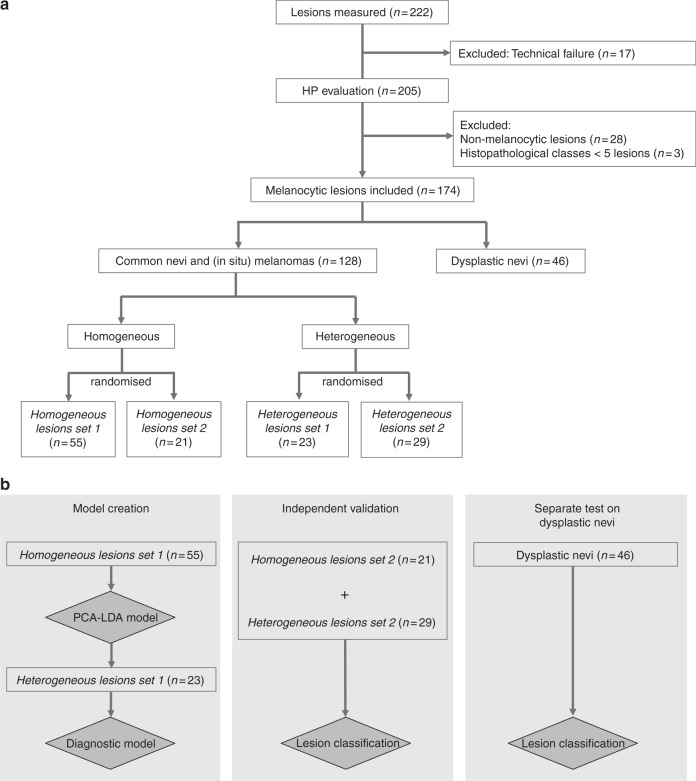
Methods: We have developed a method to improve diagnosis of (thin) melanoma, based on Raman spectroscopy. In an ex vivo study in a tertiary referral (pigmented lesions) centre, high-wavenumber Raman spectra were collected from 174 freshly excised melanocytic lesions suspicious for melanoma. Measurements were performed on multiple locations within the lesions. A diagnostic model was developed and validated on an independent data set of 96 lesions.
Results: Approximately 60% of the melanomas included in this study were melanomas in situ. The invasive melanomas had an average Breslow thickness of 0.89 mm. The diagnostic model correctly classified all melanomas (including in situ) with a specificity of 43.8%, and showed a potential improvement of the number needed to treat from 6.0 to 2.7, at a sensitivity of 100%.
Conclusion: This work signifies an important step towards accurate and objective clinical diagnosis of melanoma and in particular melanoma with Breslow thickness <0.8 mm.
Conflict of interest statement
The authors declare no competing interests.
Figures
Comment in
-
Comment on "Improving clinical diagnosis of early-stage cutaneous melanoma based on Raman spectroscopy".Br J Cancer. 2019 Apr;120(8):864. doi: 10.1038/s41416-019-0430-9. Epub 2019 Mar 22. Br J Cancer. 2019. PMID: 30899087 Free PMC article. No abstract available.
-
Reply to Comment on "Improving clinical diagnosis of early-stage cutaneous melanoma based on Raman spectroscopy".Br J Cancer. 2019 Apr;120(8):865-866. doi: 10.1038/s41416-019-0431-8. Epub 2019 Mar 22. Br J Cancer. 2019. PMID: 30899089 Free PMC article. No abstract available.
Similar articles
-
Role of In Vivo Reflectance Confocal Microscopy in the Analysis of Melanocytic Lesions.Acta Dermatovenerol Croat. 2018 Apr;26(1):64-67. Acta Dermatovenerol Croat. 2018. PMID: 29782304 Review.
-
Availability of digital dermoscopy in daily practice dramatically reduces the number of excised melanocytic lesions: results from an observational study.Br J Dermatol. 2012 Oct;167(4):778-86. doi: 10.1111/j.1365-2133.2012.11042.x. Epub 2012 Aug 20. Br J Dermatol. 2012. PMID: 22564185
-
Melanoma detection in Italian pigmented lesion clinics.G Ital Dermatol Venereol. 2014 Apr;149(2):161-6. G Ital Dermatol Venereol. 2014. PMID: 24819635
-
Assessment of Raman Spectroscopy for Reducing Unnecessary Biopsies for Melanoma Screening.Molecules. 2020 Jun 20;25(12):2852. doi: 10.3390/molecules25122852. Molecules. 2020. PMID: 32575717 Free PMC article.
-
[Strategies for the noninvasive diagnosis of melanoma].Hautarzt. 2016 Jul;67(7):519-28. doi: 10.1007/s00105-016-3796-0. Hautarzt. 2016. PMID: 27193101 Review. German.
Cited by
-
Portable System for In-Clinic Differentiation of Skin Cancers from Benign Skin Lesions and Inflammatory Dermatoses.JID Innov. 2023 Sep 29;4(1):100238. doi: 10.1016/j.xjidi.2023.100238. eCollection 2024 Jan. JID Innov. 2023. PMID: 38274304 Free PMC article.
-
Comment on "Improving clinical diagnosis of early-stage cutaneous melanoma based on Raman spectroscopy".Br J Cancer. 2019 Apr;120(8):864. doi: 10.1038/s41416-019-0430-9. Epub 2019 Mar 22. Br J Cancer. 2019. PMID: 30899087 Free PMC article. No abstract available.
-
Melanoma Biomarkers and Their Potential Application for In Vivo Diagnostic Imaging Modalities.Int J Mol Sci. 2020 Dec 16;21(24):9583. doi: 10.3390/ijms21249583. Int J Mol Sci. 2020. PMID: 33339193 Free PMC article. Review.
-
Machine Learning of Raman Spectroscopy Data for Classifying Cancers: A Review of the Recent Literature.Diagnostics (Basel). 2022 Jun 17;12(6):1491. doi: 10.3390/diagnostics12061491. Diagnostics (Basel). 2022. PMID: 35741300 Free PMC article. Review.
-
Intraoperative use of high-speed Raman spectroscopy during soft tissue sarcoma resection.Sci Rep. 2025 Mar 14;15(1):8789. doi: 10.1038/s41598-025-93089-z. Sci Rep. 2025. PMID: 40082570 Free PMC article.
References
MeSH terms
LinkOut - more resources
Full Text Sources
Medical
