. 2018 Nov 8;8(1):16568.
doi: 10.1038/s41598-018-34634-x.

Random Neuronal Networks show homeostatic regulation of global activity while showing persistent changes in specific connectivity paths to theta burst stimuli

Affiliations

Random Neuronal Networks show homeostatic regulation of global activity while showing persistent changes in specific connectivity paths to theta burst stimuli

Jude Baby George et al. Sci Rep. .

Abstract

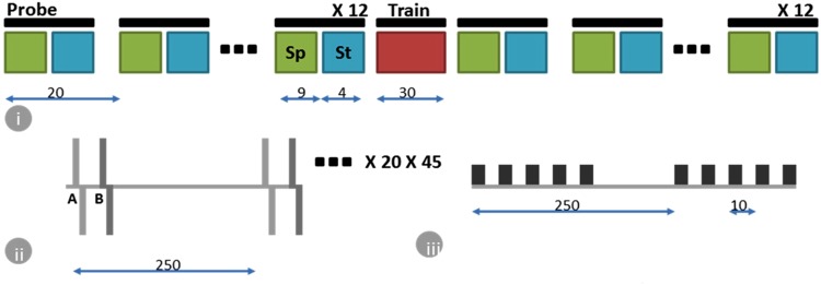
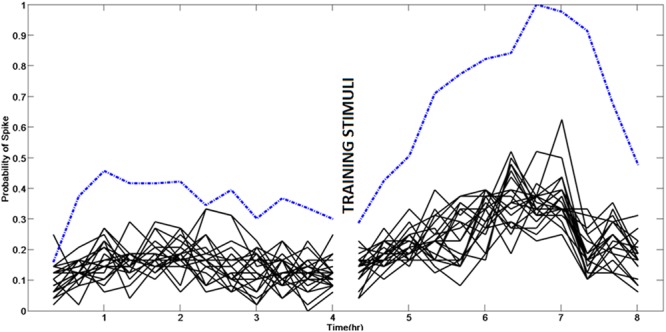
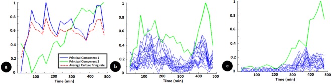
Learning in neuronal networks based on Hebbian principle has been shown to lead to destabilizing effects. Mechanisms have been identified that maintain homeostasis in such networks. However, the way in which these two opposing forces operate to support learning while maintaining stability is an active area of research. In this study, using neuronal networks grown on multi electrode arrays, we show that theta burst stimuli lead to persistent changes in functional connectivity along specific paths while the network maintains a global homeostasis. Simultaneous observations of spontaneous activity and stimulus evoked responses over several hours with theta burst training stimuli shows that global activity of the network quantified from spontaneous activity, which is disturbed due to theta burst stimuli is restored by homeostatic mechanisms while stimulus evoked changes in specific connectivity paths retain a memory trace of the training.


Conflict of interest statement

The authors declare no competing interests.

Figures

Figure 1
Figure 1
Figure shows the details of the experimental protocol in an 8.5 hr session on a day. (i) 12 monitoring sequences, each separated by 20 min, capture the state of the neuronal network before applying a training sequence (Train) for 30 min. This is followed by another set of 12 monitoring sequences to monitor the evolution of the network activity in a 4 hr window after training. Each monitoring sequence consists of a 9 min recording of spontaneous activity (Sp) and 4 min recording of stimulus evoked response (St) using probe sequences consisting of spatio-temporal stimulus patterns. This allows us to investigate, both the ongoing spontaneous activity in the network, as well as response of the network to stimuli during the same period. (ii) The spatio-temporal stimulus pattern consists of paired stimulus at 2 electrodes A and B (from a possible set of 8). The stimulus waveform at each electrode is a 500 µV biphasic voltage pulse with each phase lasting 500 µs. The time delay between end of first pulse and start of next is 500 µs. The time delay between the paired stimulus at the two electrodes is one of 0.5 ms or 3 ms. A probe sequence consists of applying 20 different spatio-temporal stimulus patterns (from a possible set of 56), applied 45 times, and each application has the 20 patterns in a random order. A delay of 250 ms is used between each stimulus pattern. (iii) The training sequence consists of repeated stimulation using one of the spatio-temporal stimulus pattern at high frequency (10 ms/100 Hz) (theta burst stimulation), repeated every 250 ms for 30 min.
Figure 2
Figure 2
Response to a specific spatio-temporal stimuli changes over time. Each circle shows spatio-temporal response of the network recorded from 120 electrodes to a specific stimulus pattern over an 8.5 hr window at times indicated. The responses shown are obtained from analyzing the outputs from probe sequences that are 60 min apart. The intensity of each dot indicates the probability of observing a spike at an output electrode, in response to that specific spatio-temporal stimuli. It can be seen that at some electrodes, post training, the probability of a spike increases while for others it decreases.
Figure 3
Figure 3
The probe response following stimulation with a training pattern at a particular output electrode to different probe patterns (black lines) is correlated with the spontaneous activity at that electrode (dashed blue line). Thus spontaneous activity recorded at an electrode could indicate its functional connectivity and current ability to respond to input stimuli. The spontaneous activity at 120 such electrodes vary differently. From this activity, we define state of the network in lower dimensions which could more concisely capture the overall network behavior and thus allow prediction of spontaneous activity and thereby response to inputs.
Figure 4
Figure 4
(a) Principle component analysis (PCA) of spontaneous activity evolution at multiple electrodes in the multi-electrode array. The solid lines indicate the first two principal components and the dashed red line indicates the average culture firing rate. (b,c) Spontaneous activity (green) and stimulus evoked responses (blue lines with different probe stimuli) at two different electrodes from the same multi-electrode dish used in (a). This shows how spontaneous activity at individual electrodes is reflected in the stimulus evoked responses. In (b), the spontaneous activity and the stimulus evoked response correlate and it is largely the reflection of the first principal component shown in a. In (c), the same correlation is observed between stimulus evoked and spontaneous activity but this largely reflects the second principal component shown in (a). It also shows how spontaneous activity at individual electrodes is different from the overall network average but can be explained by a linear combination of principal components.
Figure 5
Figure 5
Homeostatic stability and plasticity at network and single neuronal levels following training stimuli. (a) Trace of three principal components of spontaneous network activity (blue: PC1, green: PC2, red: PC3). This shows that training protocol induces a temporary disturbance in state of the network which is restored after some time. (b) Change in probability of response (pM) to specific stimuli at two different electrodes. One electrode (Green) indicates a definite increase in probability of spike response while another (Blue) indicates a persistent decrease. (c) Average culture activity during the period of recording (Spontaneous activity at each electrode is normalized and the mean for all the electrodes is plotted). There is a gradual decrease in average culture activity over the course of the experiment which shows an evolving network. But, this is not significantly affected by training protocol. This is more relevant when one observes that there is a definite disturbance in the principal components extracted from spontaneous activity which shows that pattern of activity is disturbed while overall average being same. (d) Spontaneous firing rate at electrodes (sM) indicated in b with same color. Though there is a persistent shift in evoked firing due to training, it is not so significant in spontaneous firing rate which largely follows the overall network activity as in c.

References

    1. Turrigiano G. Homeostatic Synaptic Plasticity: Local and Global Mechanisms for Stabilizing Neuronal Function. Cold Spring Harbor Perspectives in Biology. 2011;4(1):a005736–a005736. - PMC - PubMed
    1. Cruikshank Scott J, Weinberger Norman M. Evidence for the Hebbian hypothesis in experience-dependent physiological plasticity of neocortex: a critical review. Brain Research Reviews. 1996;22(3):191–228. doi: 10.1016/S0165-0173(96)00015-X. - DOI - PubMed
    1. Markram H, Lubke J, Frotscher M, Sakmann B. Regulation of synaptic efficacy by coincidence of postsynaptic APs and EPSPs. Science. 1997;275:213–215. doi: 10.1126/science.275.5297.213. - DOI - PubMed
    1. Abbott, L. F. & Nelson, S. B. Synaptic plasticity: Taming the beast. Nat Neurosci. 3, 1178–1183 (2000). - PubMed
    1. Miller Kenneth D., MacKay David J. C. The Role of Constraints in Hebbian Learning. Neural Computation. 1994;6(1):100–126. doi: 10.1162/neco.1994.6.1.100. - DOI

Publication types