. 2018 Oct 25:9:2399.
doi: 10.3389/fimmu.2018.02399. eCollection 2018.

A Natural Variant of the Signaling Molecule Vav1 Enhances Susceptibility to Myasthenia Gravis and Influences the T Cell Receptor Repertoire

Affiliations

A Natural Variant of the Signaling Molecule Vav1 Enhances Susceptibility to Myasthenia Gravis and Influences the T Cell Receptor Repertoire

Isabelle Bernard et al. Front Immunol. .

Abstract

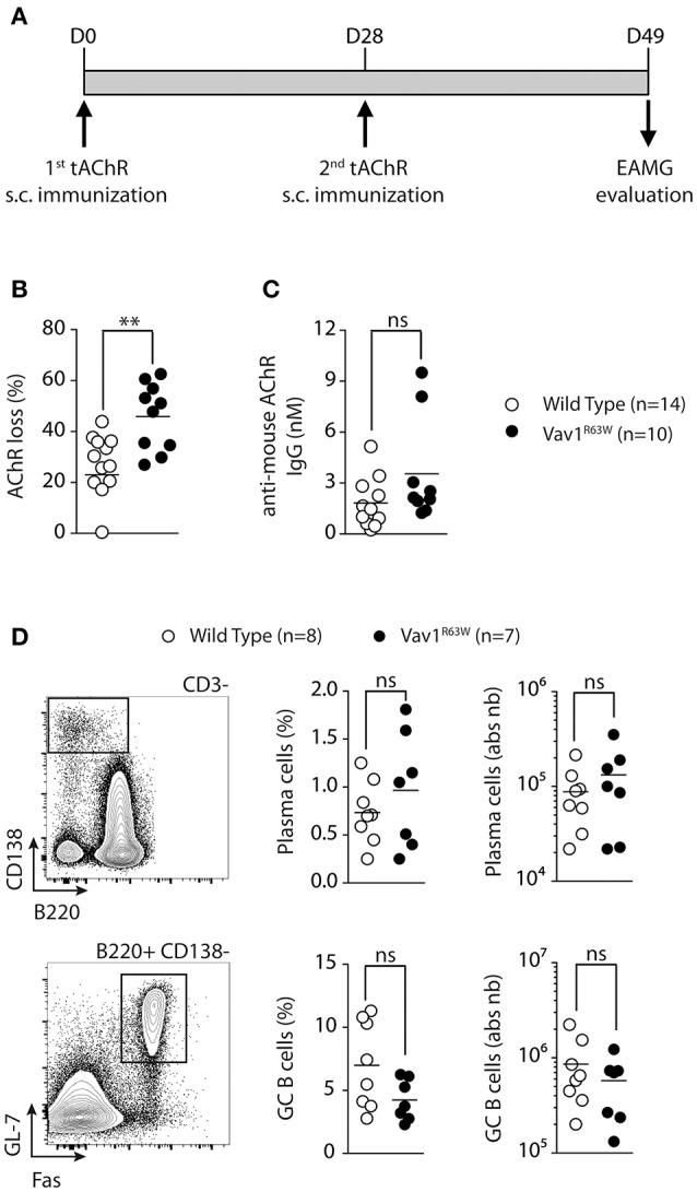
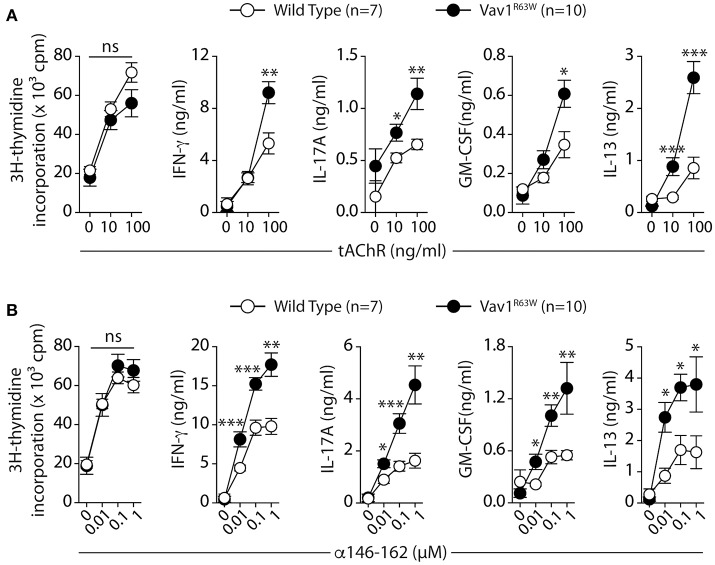
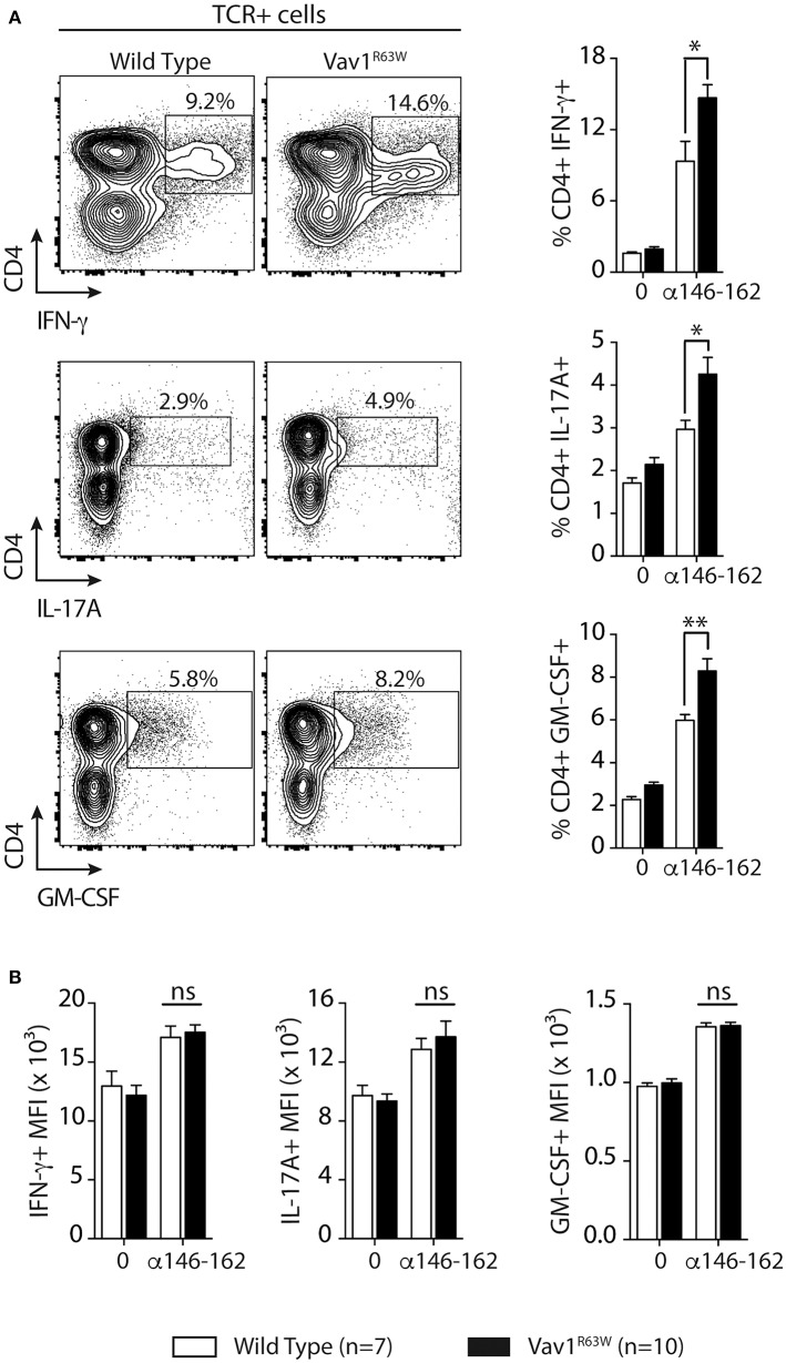
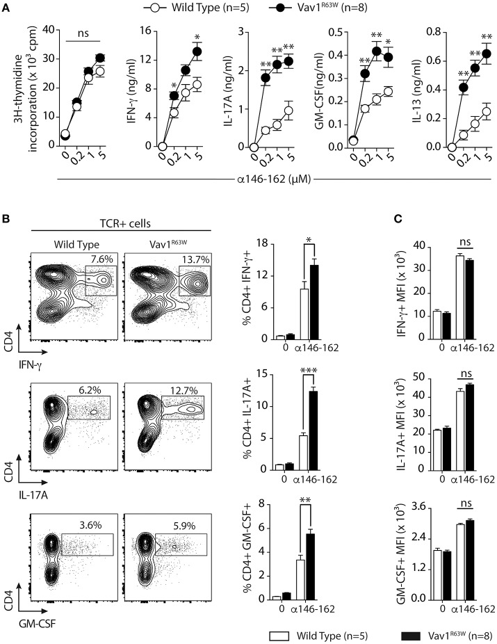
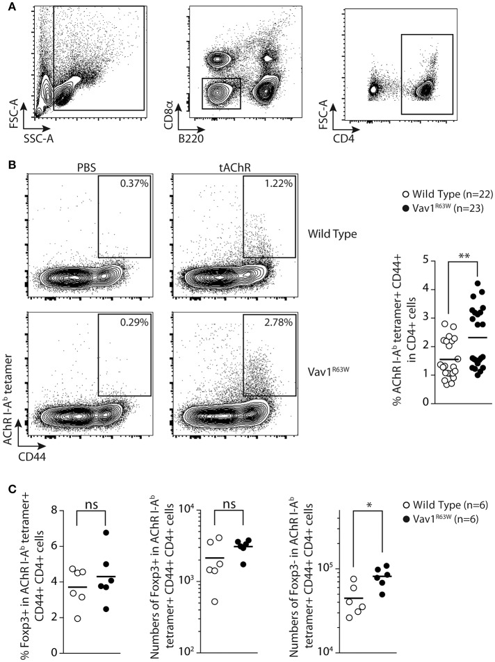
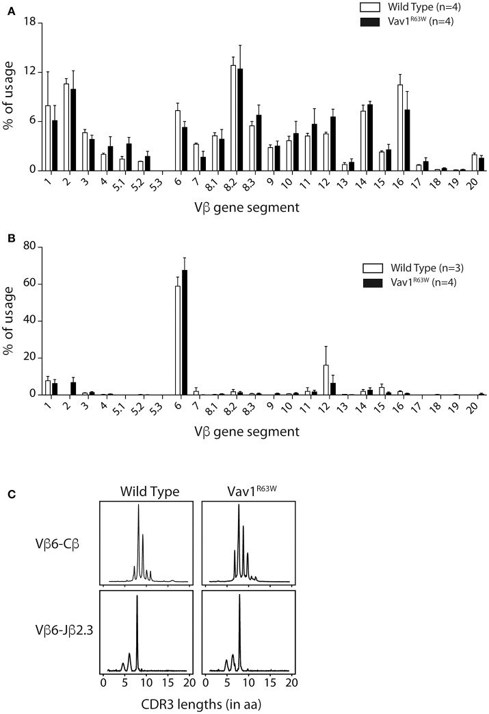
The guanine nucleotide exchange factor Vav1 is essential for transducing T cell receptor (TCR) signals and plays an important role in T cell development and activation. Previous genetic studies identified a natural variant of Vav1 characterized by the substitution of an arginine (R) residue by a tryptophane (W) at position 63 (Vav1R63W). This variant impacts Vav1 adaptor functions and controls susceptibility to T cell-mediated neuroinflammation. To assess the implication of this Vav1 variant on the susceptibility to antibody-mediated diseases, we used the animal model of myasthenia gravis, experimental autoimmune myasthenia gravis (EAMG). To this end, we generated a knock-in (KI) mouse model bearing a R to W substitution in the Vav1 gene (Vav1R63W) and immunized it with either torpedo acetylcholine receptor (tAChR) or the α146-162 immunodominant peptide. We observed that the Vav1R63W conferred increased susceptibility to EAMG, revealed by a higher AChR loss together with an increased production of effector cytokines (IFN-γ, IL-17A, GM-CSF) by antigen-specific CD4+ T cells, as well as an increased frequency of antigen-specific CD4+ T cells. This correlated with the emergence of a dominant antigen-specific T cell clone in KI mice that was not present in wild-type mice, suggesting an impact on thymic selection and/or a different clonal selection threshold following antigen encounter. Our results highlight the key role of Vav1 in the pathophysiology of EAMG and this was associated with an impact on the TCR repertoire of AChR reactive T lymphocytes.

Keywords: T cell repertoire; T cells; Vav1; animal models; myasthenia gravis.


Figures

Figure 1
Figure 1
The Vav1R63W polymorphism increases EAMG severity. EAMG was induced wild-type (WT) or Vav1R63W knock-in (Vav1R63W) mice by two immunizations with 10 μg of torpedo AChR in CFA at d0 and d28 (A). Twenty-one days after the second immunization, mice were killed for the quantification of endogenous AChR content on whole body musculature (B) and for the determination of seric concentration of IgG Abs reactive with mouse AChR (C). In (B), data were expressed as the percentage of the AChR loss calculated by considering AChR content of CFA-immunized, age-matched, control mice as 100%. Data in (B,C) represent a pool of 2 independent experiments among four performed. (D), Percentages and total cell count numbers of plasma cells and germinal center B cells determined by flow cytometry in draining LN cells of WT and Vav1R63W mice at day 9 post-tAChR immunization. The gating strategy of these two populations is depicted in (D). Each dot represents an individual mouse. Mann-Whitney test, ns, non-significant; **P < 0.01.
Figure 2
Figure 2
Production of cytokines after tAChR immunization of WT and Vav1R63W mice. WT or Vav1R63W mice were immunized with 10 μg of tAChR in CFA. Draining LNC were harvested 9 days later and stimulated for 48 h with variable concentrations of tAChR (A) or with the α146-162 immunodominant AChR peptide (B). Cytokine secretion was quantified by ELISA and CBA kit in the supernatants. Data represent a pool of 2 independent experiments. Mann-Whitney test, ns, non-significant; *P < 0.05; **P < 0.01; ***P < 0.005.
Figure 3
Figure 3
Frequency of CD4+ T cells producing cytokines after tAChR immunization of WT and Vav1R63W mice. WT or Vav1R63W mice were immunized with 10 μg of tAChR in CFA. Draining LNC were harvested 9 days later and stimulated with the α146-162 immunodominant AChR peptide. (A) Frequency of TCR+CD4+ LN cells producing cytokines was determined by intracytoplasmic staining after 72 h stimulation with (1 μM) or without α146-162 AChR peptide. (B) Histograms represent the Mean Fluorescence Intensity (MFI) of IFN-γ, IL-17A and GM-CSF expression by TCR+CD4+ LN cells. Data represent a pool of 2 independent experiments. Mann-Whitney test, ns, non-significant; *P < 0.05; **P < 0.01.
Figure 4
Figure 4
Cytokine production and frequency of CD4 T cell-producing cytokines after α146-162 AChR peptide immunization of WT and Vav1R63W mice. WT or Vav1R63W mice were immunized with 50 μg of α146-162 AChR peptide. Draining LN cells were harvested 9 days later and stimulated for 48 h with increasing concentrations of α146-162 AChR peptide. Cytokine secretion was quantified by ELISA and CBA kit in the supernatants (A). The frequency of TCR+CD4+ LN cells producing cytokines was determined by intracytoplasmic staining after 72 h stimulation without or with α146-162 AChR peptide (1 μM) (B). The intensity of expression of each cytokine by TCR+CD4+ LN cells was quantified by the Mean Fluorescence Intensity (MFI) (C). Data represent a pool of 2 independent experiments. Mann-Whitney test, ns, non-significant; *P < 0.05; **P < 0.01; ***P < 0.005.
Figure 5
Figure 5
Tracking AChR-specific CD4+ T cells in WT and Vav1R63W KI mice. Nine days after sub-cutaneous immunization, draining LN were harvested and analyzed by flow cytometry. The gating strategy of total CD4+ T cells is depicted in (A) by excluding B220+ and CD8α+ cells and by positively selecting CD4+ cells. The detection of tAChR-specific activated CD4+ T cells (AChR I-Ab tetramer+ CD44+) are depicted for wild-type and Vav1R63W KI mice after immunization with PBS in CFA or tAChR in CFA (B) (n = 22 for wild-type mice and n = 23 for Vav1R63W KI mice). Data represent a pool of 4 independent experiments. Intracellular expression of Foxp3 was assessed in tAChR-specific CD4+ T cells. Frequency of Foxp3+ Treg and absolute numbers of Foxp3+ Treg and Foxp3- effector tAChR-specific CD4+ T cells are depicted (C) (n = 6 for wild-type mice and n = 6 for Vav1R63W KI mice). Each dot represents an individual mouse; horizontal lines denote the mean value of groups. Mann-Whitney test, *P < 0.05, **P < 0.01.
Figure 6
Figure 6
Vß gene segment usage and CDR3 size distribution of α146-162 AChR-specific CD4+ T cells from WT and Vav1R63W mice. Nine days after sub-cutaneous immunization, draining LN were harvested and tAChR-specific activated CD4+ T cells (AChR I-Ab tetramer+ CD44+) were FACS sorted out prior to RNA isolation and RT-coupled real time PCR analysis. Alternatively, lymph node CD4+ T cells were isolated from naive mice. Vβ gene usage for naive (A) and tAChR-specific CD4+ T cells (B) isolated from WT (white) or Vav1R63W (black) mice are depicted. CDR3b size distributions from one representative sample of each background are presented (C). Rearrangements shown are Vß6-Cß and Vß6-Jα2.3.

References

    1. Vincent A. Unravelling the pathogenesis of myasthenia gravis. Nat Rev Immunol. (2002) 2:797–804. 10.1038/nri916 - DOI - PubMed
    1. Berrih-Aknin S, Frenkian-Cuvelier M, Eymard B. Diagnostic and clinical classification of autoimmune myasthenia gravis. J Autoimmun. (2014) 48–49:143–8. 10.1016/j.jaut.2014.01.003 - DOI - PubMed
    1. Phillips WD, Vincent A. Pathogenesis of myasthenia gravis: update on disease types, models, and mechanisms. F1000Res (2016) 5:1–10 10.12688/f1000research.8206.1 - DOI - PMC - PubMed
    1. Kordas G, Lagoumintzis G, Sideris S, Poulas K, Tzartos SJ. Direct proof of the in vivo pathogenic role of the AChR autoantibodies from myasthenia gravis patients. PLoS ONE (2014) 9:e108327. 10.1371/journal.pone.0108327 - DOI - PMC - PubMed
    1. Hoch W, McConville J, Helms S, Newsom-Davis J, Melms A, Vincent A. Auto-antibodies to the receptor tyrosine kinase MuSK in patients with myasthenia gravis without acetylcholine receptor antibodies. Nat Med. (2001) 7:365–8. 10.1038/85520 - DOI - PubMed

Publication types

MeSH terms