Review
. 2018 Oct 25:9:2469.
doi: 10.3389/fimmu.2018.02469. eCollection 2018.

Regulation of the Germinal Center Response

Affiliations
Review

Regulation of the Germinal Center Response

Marisa Stebegg et al. Front Immunol. .

Abstract

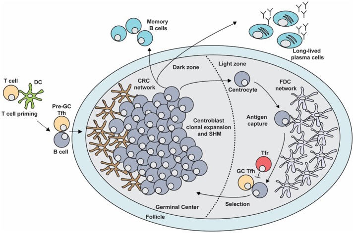
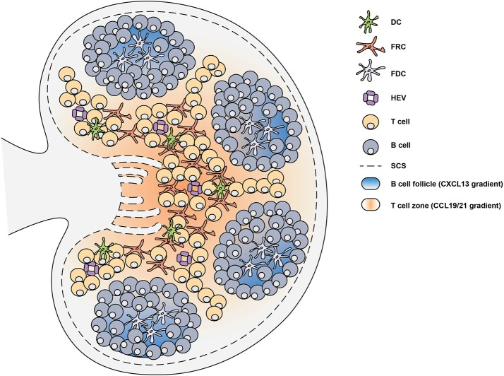
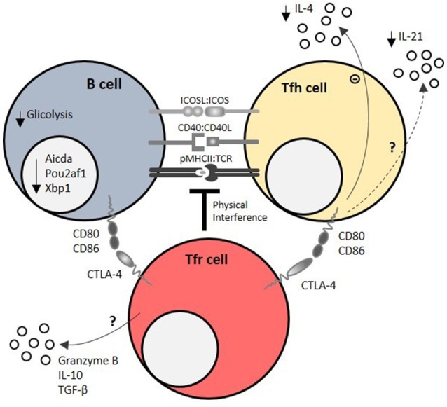
The germinal center (GC) is a specialized microstructure that forms in secondary lymphoid tissues, producing long-lived antibody secreting plasma cells and memory B cells, which can provide protection against reinfection. Within the GC, B cells undergo somatic mutation of the genes encoding their B cell receptors which, following successful selection, can lead to the emergence of B cell clones that bind antigen with high affinity. However, this mutation process can also be dangerous, as it can create autoreactive clones that can cause autoimmunity. Because of this, regulation of GC reactions is critical to ensure high affinity antibody production and to enforce self-tolerance by avoiding emergence of autoreactive B cell clones. A productive GC response requires the collaboration of multiple cell types. The stromal cell network orchestrates GC cell dynamics by controlling antigen delivery and cell trafficking. T follicular helper (Tfh) cells provide specialized help to GC B cells through cognate T-B cell interactions while Foxp3+ T follicular regulatory (Tfr) cells are key mediators of GC regulation. However, regulation of GC responses is not a simple outcome of Tfh/Tfr balance, but also involves the contribution of other cell types to modulate the GC microenvironment and to avoid autoimmunity. Thus, the regulation of the GC is complex, and occurs at multiple levels. In this review we outline recent developments in the biology of cell subsets involved in the regulation of GC reactions, in both secondary lymphoid tissues, and Peyer's patches (PPs). We discuss the mechanisms which enable the generation of potent protective humoral immunity whilst GC-derived autoimmunity is avoided.

Keywords: Tfh cell; Tfr cell; germinal center (GC); humoral responses; immuneregulation.


Figures

Figure 1
Figure 1
The germinal center (GC) response. The GC is a specialized microenvironment formed within the B cell follicles of secondary lymphoid tissues upon infection or immunization. The GC is divided into two distinct compartments. The dark zone (DZ) that contains a network of CXCL12-producing reticular cells (CRCs) and is the site of GC B cell proliferation and somatic hypermutation (SHM). Centroblasts then follow a CXCL13 gradient to enter the light zone (LZ) as centrocytes through their expression of CXCR5. In the LZ, centrocytes capture antigen presented on follicular dendritic cells (FDCs) which they internalize, process and subsequently present to T follicular helper (Tfh) cells in order to undergo selection. This process is regulated by T follicular regulatory (Tfr) cells which are also present in the LZ. Upon receiving survival signals from Tfh cells, centrocytes re-enter the DZ for further rounds of proliferation and SHM after which they exit the GC as memory B cells or high-affinity antibody-secreting plasma cells.
Figure 2
Figure 2
Lymph node structure is supported by stromal cells. Secondary lymphoid organs are divided into distinct regions through the generation of chemokine gradients by stromal cells. In the lymph node (LN), these chemokine gradients allow the circulation of lymphocytes which enter through high endothelial venules (HEV). In the T cell zone, fibroblastic reticular cells (FRCs) generate a CCL19 and CCL21 gradient which facilitates the migration of T cells and dendritic cells (DCs). The B cell follicles contain follicular dendritic cells (FDCs) which generate a CXCL13 gradient that promote the localization of B cells. Upon infection or immunization, antigen can enter the LN through the subcapsular sinus (SCS) or can be brought by DCs to trigger the activation of lymphocytes.
Figure 3
Figure 3
Mechanisms of Tfr cell-mediated regulation of humoral responses. Tfr cells regulate T – B interactions within the germinal center (GC) by physical interference at the immunological synapse, which is required for the survival feedback loop between GC B cells and T follicular helper (Tfh) cells. CTLA-4 is a key molecule of T follicular regulatory (Tfr) cell function at immunological synapse, as it directly blocks CD80/CD86 co-stimulatory signals. Using these mechanisms, Tfr cells impair GC B cell metabolism (mainly by decreasing glucose uptake and usage) and induce the downregulation of GC B cell effector molecules, such as Pou2af1 (required for GC B cell formation), Xbp1 (required for antibody secretion), and Aicda (required for class switch recombination). On the other side of the immunological synapse, Tfr cells limit IL-21, and IL-4 secretion by Tfh cells. Granzyme B, IL-10 and TGF-β secretion by Tfr cells may also account for their regulatory capacity.

References

    1. MacLennan ICM. Germinal centers. Annu Rev Immunol. (1994) 12:117–39. 10.1146/annurev.iy.12.040194.001001 - DOI - PubMed
    1. Pitzalis C, Jones GW, Bombardieri M, Jones SA. Ectopic lymphoid-like structures in infection, cancer and autoimmunity. Nat Rev Immunol. (2014) 14:447–62. 10.1038/nri3700 - DOI - PubMed
    1. Mesin L, Ersching J, Victora GD. Germinal center B cell dynamics. Immunity (2016) 45:471–82. 10.1016/j.immuni.2016.09.001 - DOI - PMC - PubMed
    1. Allen CDC, Cyster JG. Follicular dendritic cell networks of primary follicles and germinal centers: phenotype and function. Semin Immunol. (2008) 20:14–25. 10.1016/j.smim.2007.12.001 - DOI - PMC - PubMed
    1. Batista FD, Harwood NE. The who, how and where of antigen presentation to B cells. Nat Rev Immunol. (2009) 9:15–27. 10.1038/nri2454 - DOI - PubMed

Publication types