. 2018 Oct 31;3(10):12392-12402.
doi: 10.1021/acsomega.8b01451. Epub 2018 Oct 1.

Ivermectin Promotes Peripheral Nerve Regeneration during Wound Healing

Affiliations

Ivermectin Promotes Peripheral Nerve Regeneration during Wound Healing

Dana M Cairns et al. ACS Omega. .

Abstract

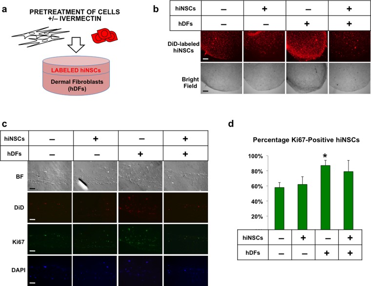
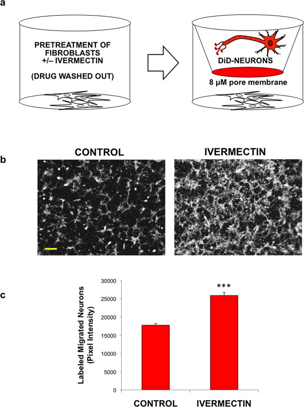
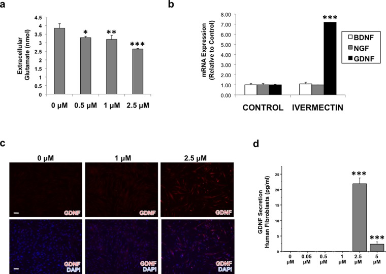
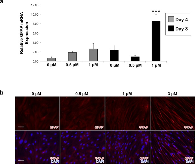
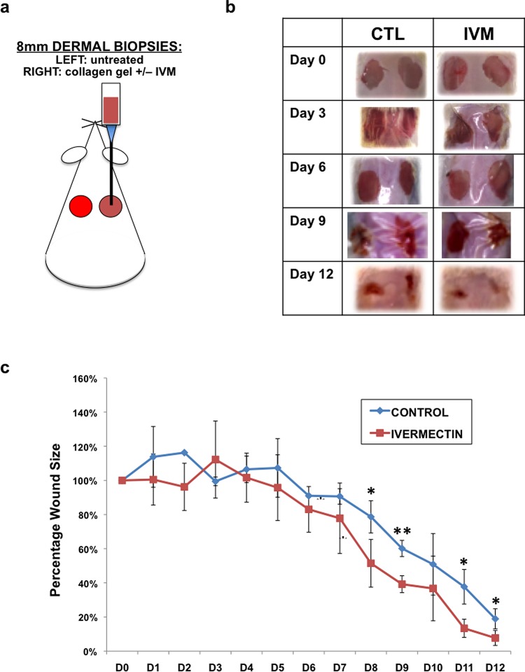
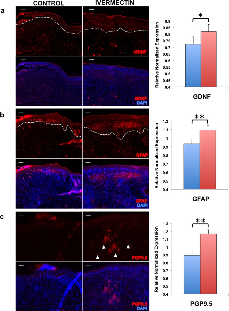
Peripheral nerves have the capacity to regenerate due to the presence of neuroprotective glia of the peripheral nervous system, Schwann cells. Upon peripheral nerve injury, Schwann cells create a permissive microenvironment for neuronal regrowth by taking up cytotoxic glutamate and secreting neurotrophic signaling molecules. Impaired peripheral nerve repair is often caused by a defective Schwann cell response after injury, and there is a critical need to develop new strategies to enhance nerve regeneration, especially in organisms with restricted regenerative potential, such as humans. One approach is to explore mechanisms in lower organisms, in which nerve repair is much more efficient. A recent study demonstrated that the antiparasitic drug, ivermectin, caused hyperinnervation of primordial eye tissue in Xenopus laevis tadpoles. Our study aimed to examine the role of ivermectin in mammalian nerve repair. We performed in vitro assays utilizing human induced neural stem cells (hiNSCs) in co-culture with human dermal fibroblasts (hDFs) and found that ivermectin-treated hDFs promote hiNSC proliferation and migration. We also characterized the effects of ivermectin on hDFs and found that ivermectin causes hDFs to uptake extracellular glutamate, secrete glial cell-derived neurotrophic factor, develop an elongated bipolar morphology, and express glial fibrillary acidic protein. Finally, in a corresponding in vivo model, we found that localized ivermectin treatment in a dermal wound site induced the upregulation of both glial and neuronal markers upon healing. Taken together, we demonstrate that ivermectin promotes peripheral nerve regeneration by inducing fibroblasts to adopt a glia-like phenotype.


Conflict of interest statement

The authors declare no competing financial interest.

Figures

Figure 1
Figure 1
Treatment of dermal fibroblasts with ivermectin induces proliferation in adjacent neural stem cells in 3D co-cultures. (a) Schematic diagram of experimental design. Human dermal fibroblasts (hDFs) and human induced neural stem cells (hiNSCs) fluorescently labeled with DiD dye were separately treated with or without 1 μM ivermectin (as indicated by “+” or “–”, respectively) and subsequently washed repeatedly to remove the drug, seeded into 3D bilayer collagen gel constructs, and cultured for 5 days. (b) Low-magnification view of 3D collagen gel constructs, scale bar: 500 μM. (c) Cryosections of collagen gels immunostained for proliferation marker, Ki67, scale bar: 100 μM. (d) Quantification of Ki67-positive DiD-labeled neural stem cells. *P ≤ 0.05, **P ≤ 0.01, ***P ≤ 0.001; as determined by one-way analysis of variance (ANOVA) with post-hoc Tukey test. Error bars show mean ± SD.
Figure 2
Figure 2
Treatment of dermal fibroblasts with ivermectin induces migration of differentiated neurons. (a) Schematic diagram of experimental design. Human dermal fibroblasts were seeded into the bottom of cell culture plates, subsequently treated with or without ivermectin, and washed repeatedly to remove the drug. Differentiated DiD-labeled neurons were seeded onto coated transwells (8 μM pore size), which were placed into the wells containing fibroblasts. Cells were cultured in low serum media (to minimize potential cell proliferation) overnight, and the relative number of cells migrating to the bottom of transwells was quantified. (b) Images of fluorescently labeled neurons that migrated to the bottom of transwells upon co-culture with dermal fibroblasts pretreated with or without ivermectin, scale bar: 200 μM. (c) Quantification of migrated cells. *P ≤ 0.05, **P ≤ 0.01, ***P ≤ 0.001; as determined by two-tailed t-test. Error bars show mean ± SD.
Figure 3
Figure 3
Treatment with ivermectin causes dermal fibroblasts to uptake extracellular glutamate and to express glial cell line-derived neurotrophic growth factor (GDNF). (a) Dermal fibroblasts were treated with various concentrations of ivermectin overnight, and cell culture media was assayed to determine extracellular glutamate concentration. (b) Dermal fibroblasts were treated with or without 1 μM ivermectin for 4 days, then subjected to quantitative real-time polymerase chain reaction (qRT-PCR) analysis for various neurotrophic growth factors. (c) Immunostaining results of dermal fibroblasts treated with ivermectin show an increase in GDNF expression with increasing ivermectin concentration, scale bar: 100 μM. (d) Enzyme-linked immunosorbent assay (ELISA) of cell culture media harvested from dermal fibroblasts treated with ivermectin for 4 days indicates that GDNF is secreted from ivermectin-treated fibroblasts. *P ≤ 0.05, **P ≤ 0.01, ***P ≤ 0.001; as determined by one-way ANOVA with post-hoc Tukey test. Error bars show mean ± SD.
Figure 4
Figure 4
Treatment of dermal fibroblasts with increasing concentrations of ivermectin results in the upregulation of GFAP as well as the development of an elongated morphology reminiscent of Schwann cells. (a) Dermal fibroblasts were treated with varying concentrations of ivermectin for 4 and 8 days, then subjected to qRT-PCR analysis of GFAP expression. (b) GFAP immunostaining demonstrates that dermal fibroblasts treated with relatively higher concentrations of ivermectin for 8 days results in an increase of GFAP expression as well as a change in morphology, which resembles a Schwann cell-like phenotype, scale bar: 100 μM. ***P ≤ 0.001; as determined by one-way ANOVA with post-hoc Tukey test. Error bars show mean ± SD.
Figure 5
Figure 5
Ivermectin promotes wound healing of dermal biopsies in vivo. (a) Schematic diagram of experimental design. Biopsies (2 × 8 mm2) were taken from the dorsal dermal layer of each mouse. In the right side wound, 30 μL collagen gels containing 10 μM ivermectin or DMSO (control) were pipetted onto the wound and allowed to solidify. The left side wounds remained untreated, and served as additional controls. Both wounds were sealed using Tegaderm, and wound progression was followed over the course of 12 days. (b) Images of gross morphology of wound healing over time. (c) Quantification of wound size over time. *P ≤ 0.05, **P ≤ 0.01; as determined by two-tailed t-test. Error bars show mean ± SD.
Figure 6
Figure 6
Ivermectin facilitates wound healing by inducing the differentiation of glia-like cells that promote nerve growth. Cryosections of the wound sites were immunostained and quantified to assay for the presence of (a) glial-derived growth factor (GDNF), (b) glial fibrillary acidic protein (GFAP), and (c) peripheral nerve marker (PGP9.5), scale bar: 100 μM. *P ≤ 0.05, **P ≤ 0.01, ***P ≤ 0.001; as determined by two-tailed t-test. Error bars show mean ± SD.

References

    1. Lundborg G.; Richard P. Nerve injury and repair--a challenge to the plastic brain. J. Peripher. Nerv. Syst. 2003, 8, 209–226. 10.1111/j.1085-9489.2003.03027.x. - DOI - PubMed
    1. Martins R. S.; Bastos D.; Siqueira M. G.; Heise C. O.; Teixeira M. J. Traumatic injuries of peripheral nerves: a review with emphasis on surgical indication. Arq. Neuro-Psiquiatr. 2013, 71, 811–814. 10.1590/0004-282X20130127. - DOI - PubMed
    1. Grinsell D.; Keating C. P. Peripheral nerve reconstruction after injury: a review of clinical and experimental therapies. Biomed. Res. Int. 2014, 2014, 69825610.1155/2014/698256. - DOI - PMC - PubMed
    1. Fu S. Y.; Gordon T. Contributing factors to poor functional recovery after delayed nerve repair: prolonged denervation. J. Neurosci. 1995, 15, 3886–3895. 10.1523/JNEUROSCI.15-05-03886.1995. - DOI - PMC - PubMed
    2. Painter M. W.; Brosius Lutz A.; Cheng Y. C.; Latremoliere A.; Duong K.; Miller C. M.; Posada S.; Cobos E. J.; Zhang A. X.; Wagers A. J.; Havton L. A.; Barres B.; Omura T.; Woolf C. J. Diminished Schwann cell repair responses underlie age-associated impaired axonal regeneration. Neuron 2014, 83, 331–343. 10.1016/j.neuron.2014.06.016. - DOI - PMC - PubMed
    1. Wong K. M.; Babetto E.; Beirowski B. Axon degeneration: make the Schwann cell great again. Neural Regener. Res. 2017, 12, 518–524. 10.4103/1673-5374.205000. - DOI - PMC - PubMed