Review
. 2018 Dec 1;125(6):1851-1859.
doi: 10.1152/japplphysiol.00113.2018. Epub 2018 Nov 9.

Microvascular mechanisms limiting skeletal muscle blood flow with advancing age

Affiliations
Review

Microvascular mechanisms limiting skeletal muscle blood flow with advancing age

Matthew J Socha et al. J Appl Physiol (1985). .

Abstract

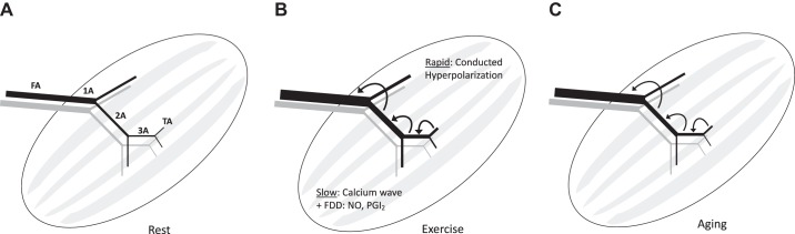
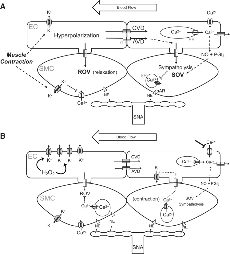
Effective oxygen delivery to active muscle fibers requires that vasodilation initiated in distal arterioles, which control flow distribution and capillary perfusion, ascends the resistance network into proximal arterioles and feed arteries, which govern total blood flow into the muscle. With exercise onset, ascending vasodilation reflects initiation and conduction of hyperpolarization along endothelium from arterioles into feed arteries. Electrical coupling of endothelial cells to smooth muscle cells evokes the rapid component of ascending vasodilation, which is sustained by ensuing release of nitric oxide during elevated luminal shear stress. Concomitant sympathetic neural activation inhibits ascending vasodilation by stimulating α-adrenoreceptors on smooth muscle cells to constrict the resistance vasculature. We hypothesized that compromised muscle blood flow in advanced age reflects impaired ascending vasodilation through actions on both cell layers of the resistance network. In the gluteus maximus muscle of old (24 mo) vs. young (4 mo) male mice (corresponding to mid-60s vs. early 20s in humans) inhibition of α-adrenoreceptors in old mice restored ascending vasodilation, whereas even minimal activation of α-adrenoreceptors in young mice attenuated ascending vasodilation in the manner seen with aging. Conduction of hyperpolarization along the endothelium is impaired in old vs. young mice because of "leaky" membranes resulting from the activation of potassium channels by hydrogen peroxide released from endothelial cells. Exposing the endothelium of young mice to hydrogen peroxide recapitulates this effect of aging. Thus enhanced α-adrenoreceptor activation of smooth muscle in concert with electrically leaky endothelium restricts muscle blood flow by impairing ascending vasodilation in advanced age.

Keywords: adrenoreceptors; aging; ascending vasodilation; endothelium; potassium channels.


Conflict of interest statement

No conflicts of interest, financial or otherwise, are declared by the authors.

Figures

Fig. 1.
Fig. 1.
Ascending vasodilation in arteriolar networks of skeletal muscle. A: schematic depiction of skeletal muscle microvascular network (black, arterial; gray, venous) beginning with the feed artery (FA) that enters the muscle and transitions into a first-order arteriole (1A). The 1A branches into smaller second-order arterioles (2A), which further branch into third-order arterioles (3A), which ultimately give rise to the terminal arterioles (TA) that control capillary perfusion (not shown). B: in response to skeletal muscle contraction, vasodilation initiated in downstream branches ascends the resistance network into the proximal FA to attain peak increases in muscle blood flow according to the metabolic demand of contracting muscle fibers. The initial rapid component of ascending vasodilation occurs in response to activation of K+ channels, resulting in hyperpolarization that is conducted upstream from cell to cell through gap junctions. The ensuing slower component of ascending vasodilation results from a calcium wave along the endothelium and flow-dependent dilation (FDD) occurring secondary to an increase in luminal shear stress that produces the vasodilator autacoids nitric oxide (NO) and prostacyclin (PGI2). C: with aging, ascending vasodilation is attenuated, thereby limiting muscle blood flow. See Fig. 2 for details of signaling events.
Fig. 2.
Fig. 2.
Microvascular mechanisms limiting skeletal muscle blood flow with advancing age. A: skeletal muscle contraction (and Ca2+ influx) activates small- and intermediate-conductance K+ channels in endothelial cells (ECs) and inward-rectifying K+ channels of smooth muscle cells (SMCs). The efflux of K+ results in hyperpolarization, thereby inhibiting Ca2+ entry into SMCs through voltage-gated Ca2+ channels and effecting rapid onset vasodilation (ROV). In the endothelium, conducted vasodilation (CVD) and ascending vasodilation (AVD) ensue as hyperpolarization spreads through gap junctions (GJs) from EC to EC and into SMCs through myoendothelial GJs. EC intracellular calcium ion concentration ([Ca2+]i) increases with internal release [from the endoplasmic reticulum (ER)], leading to a Ca2+ wave that spreads from EC to EC, complemented by Ca2+ influx through transient receptor potential channels in response to blood flow (shear stress). The rise in EC [Ca2+]i generates nitric oxide (NO) and prostacyclin (PGI2) that mediate slow onset vasodilation (SOV). These dilator signals override concurrent sympathetic nerve activity (SNA) that releases norepinephrine (NE) onto SMC, attenuating the effects of α-adrenoreceptor (αAR) activation that promote vasoconstriction through release of Ca2+ from the sarcoplasmic reticulum (SR). The net result is functional sympatholysis with elevated blood flow to active skeletal muscle fibers. B: key effects of aging. Elevated hydrogen peroxide (H2O2) in ECs activates K+ channels in the plasma membrane, thereby increasing K+ efflux and attenuating the spread of hyperpolarization and reducing CVD and AVD (as depicted in Fig. 1C) along with sympatholysis. Concurrent increases in SNA result in greater NE release onto SMCs, promoting SMC contraction; the rise in SMC [Ca2+]i promotes Ca2+ diffusion through myoendothelial GJs, further activating K+ channels in ECs to augment signal dissipation. Endothelial dysfunction attenuates the effect of shear stress, reducing the generation of NO and PGI2 and attenuating SOV. A reduction in Ca2+ entry through transient receptor potential channels has a protective effect on the endothelium during oxidative stress.

References

    1. Andersen P, Saltin B. Maximal perfusion of skeletal muscle in man. J Physiol 366: 233–249, 1985. doi:10.1113/jphysiol.1985.sp015794. - DOI - PMC - PubMed
    1. Bagher P, Segal SS. Regulation of blood flow in the microcirculation: role of conducted vasodilation. Acta Physiol (Oxf) 202: 271–284, 2011. doi:10.1111/j.1748-1716.2010.02244.x. - DOI - PMC - PubMed
    1. Bearden SE, Linn E, Ashley BS, Looft-Wilson RC. Age-related changes in conducted vasodilation: effects of exercise training and role in functional hyperemia. Am J Physiol Regul Integr Comp Physiol 293: R1717–R1721, 2007. doi:10.1152/ajpregu.00827.2006. - DOI - PubMed
    1. Bearden SE, Payne GW, Chisty A, Segal SS. Arteriolar network architecture and vasomotor function with ageing in mouse gluteus maximus muscle. J Physiol 561: 535–545, 2004. doi:10.1113/jphysiol.2004.068262. - DOI - PMC - PubMed
    1. Bearden SE, Segal SS. Neurovascular alignment in adult mouse skeletal muscles. Microcirculation 12: 161–167, 2005. doi:10.1080/10739680590904964. - DOI - PubMed

Publication types