

Elevated arterial shear rate increases indexes of endothelial cell autophagy and nitric oxide synthase activation in humans
- PMID: 30412436
- PMCID: PMC6734082
- DOI: 10.1152/ajpheart.00561.2018
Elevated arterial shear rate increases indexes of endothelial cell autophagy and nitric oxide synthase activation in humans
Abstract
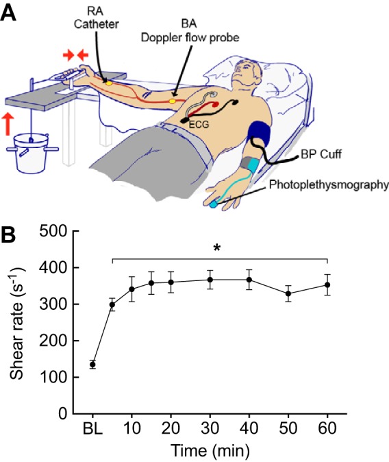
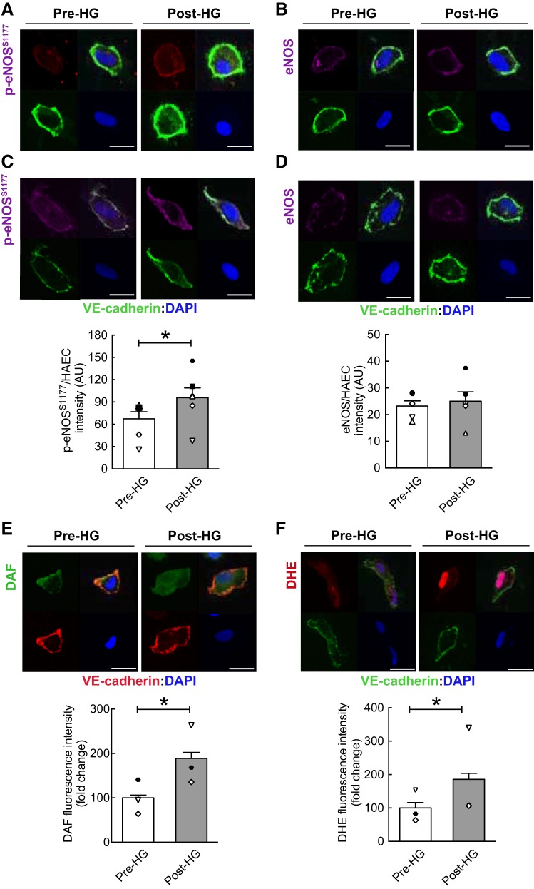
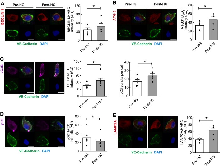
Continuous laminar shear stress increases the process of autophagy, activates endothelial nitric oxide (NO) synthase phosphorylation at serine 1177 (p-eNOSS1177), and generates NO in bovine and human arterial endothelial cells (ECs) compared with static controls. However, the translational relevance of these findings has not been explored. In the current study, primary ECs were collected from the radial artery of 7 men using sterile J-wires before (Pre) and after (Post) 60 min of rhythmic handgrip exercise (HG) performed with the same arm. After ECs were identified by positive costaining for vascular endothelial cadherin and 4',6'-diamidino-2-phenylindole, immunofluorescent antibodies were used to assess indices of autophagy, NO generation, and superoxide anion (O2·-) production. Commercially available primary human arterial ECs were stained and processed in parallel to serve as controls. All end points were evaluated using 75 ECs from each subject. Relative to Pre-HG, HG elevated arterial shear rate ( P < 0.05) ~3-fold, whereas heart rate, arterial pressure, and cardiac output were not altered. Compared with values obtained from ECs Pre-HG, Post-HG ECs displayed increased ( P < 0.05) expression of p-eNOSS1177, NO generation, O2·- production, BECLIN1, microtubule-associated proteins 1A/1B light chain 3B, autophagy-related gene 3, and lysosomal-associated membrane protein 2A and decreased ( P < 0.05) expression (i.e., enhanced degradation) of the adaptor protein p62/sequestosome-1. These novel findings provide evidence that elevated arterial shear rate associated with functional hyperemia initiates autophagy, activates p-eNOSS1177, and increases NO and O2·- generation in primary human ECs. NEW & NOTEWORTHY Previously, our group reported in bovine arterial and human arterial endothelial cells (ECs) that shear stress initiates trafficking of the autophagosome to the lysosome and increases endothelial nitric oxide (NO) synthase phosphorylation at serine 1177, NO generation, and O2·- production. Here, the translational relevance of these findings is documented. Specifically, functional hyperemia induced by rhythmic handgrip exercise elevates arterial shear rate to an extent that increases indices of autophagy, NO generation, and O2·- production in primary arterial ECs collected from healthy men.
Keywords: blood vessel; exercise; immunofluorescence; shear stress.
Conflict of interest statement
No conflicts of interest, financial or otherwise, are declared by the authors.
Figures
Comment in
-
Vascular autophagy in physiology and pathology.Am J Physiol Heart Circ Physiol. 2019 Jan 1;316(1):H183-H185. doi: 10.1152/ajpheart.00707.2018. Epub 2018 Nov 9. Am J Physiol Heart Circ Physiol. 2019. PMID: 30412440 Free PMC article. No abstract available.
References
-
- Bharath LP, Cho JM, Park SK, Ruan T, Li Y, Mueller R, Bean T, Reese V, Richardson RS, Cai J, Sargsyan A, Pires K, Anandh Babu PV, Boudina S, Graham TE, Symons JD. Endothelial cell autophagy maintains shear stress-induced nitric oxide generation via glycolysis-dependent purinergic signaling to endothelial nitric oxide synthase. Arterioscler Thromb Vasc Biol 37: 1646–1656, 2017. doi: 10.1161/ATVBAHA.117.309510. - DOI - PMC - PubMed
-
- Bharath LP, Mueller R, Li Y, Ruan T, Kunz D, Goodrich R, Mills T, Deeter L, Sargsyan A, Anandh Babu PV, Graham TE, Symons JD. Impairment of autophagy in endothelial cells prevents shear-stress-induced increases in nitric oxide bioavailability. Can J Physiol Pharmacol 92: 605–612, 2014. doi: 10.1139/cjpp-2014-0017. - DOI - PMC - PubMed
-
- Bharath LP, Ruan T, Li Y, Ravindran A, Wan X, Nhan JK, Walker ML, Deeter L, Goodrich R, Johnson E, Munday D, Mueller R, Kunz D, Jones D, Reese V, Summers SA, Babu PV, Holland WL, Zhang QJ, Abel ED, Symons JD. Ceramide-initiated protein phosphatase 2A activation contributes to arterial dysfunction in vivo. Diabetes 64: 3914–3926, 2015. doi: 10.2337/db15-0244. - DOI - PMC - PubMed
Publication types
MeSH terms
Substances
Grants and funding
LinkOut - more resources
Full Text Sources
Medical