. 2018 Nov 8;7(11):423.
doi: 10.3390/jcm7110423.

Post-Translational Modifications and Diastolic Calcium Leak Associated to the Novel RyR2-D3638A Mutation Lead to CPVT in Patient-Specific hiPSC-Derived Cardiomyocytes

Affiliations

Post-Translational Modifications and Diastolic Calcium Leak Associated to the Novel RyR2-D3638A Mutation Lead to CPVT in Patient-Specific hiPSC-Derived Cardiomyocytes

Ivana Acimovic et al. J Clin Med. .

Abstract

Background: Sarcoplasmic reticulum Ca2+ leak and post-translational modifications under stress have been implicated in catecholaminergic polymorphic ventricular tachycardia (CPVT), a highly lethal inherited arrhythmogenic disorder. Human induced pluripotent stem cells (hiPSCs) offer a unique opportunity for disease modeling.

Objective: The aims were to obtain functional hiPSC-derived cardiomyocytes from a CPVT patient harboring a novel ryanodine receptor (RyR2) mutation and model the syndrome, drug responses and investigate the molecular mechanisms associated to the CPVT syndrome.

Methods: Patient-specific cardiomyocytes were generated from a young athletic female diagnosed with CPVT. The contractile, intracellular Ca2+ handling and electrophysiological properties as well as the RyR2 macromolecular remodeling were studied.

Results: Exercise stress electrocardiography revealed polymorphic ventricular tachycardia when treated with metoprolol and marked improvement with flecainide alone. We found abnormal stress-induced contractile and electrophysiological properties associated with sarcoplasmic reticulum Ca2+ leak in CPVT hiPSC-derived cardiomyocytes. We found inadequate response to metoprolol and a potent response of flecainide. Stabilizing RyR2 with a Rycal compound prevents those abnormalities specifically in CPVT hiPSC-derived cardiomyocytes. The RyR2-D3638A mutation is located in the conformational change inducing-central core domain and leads to RyR2 macromolecular remodeling including depletion of PP2A and Calstabin2.

Conclusion: We identified a novel RyR2-D3638A mutation causing 3D conformational defects and aberrant biophysical properties associated to RyR2 macromolecular complex post-translational remodeling. The molecular remodeling is for the first time revealed using patient-specific hiPSC-derived cardiomyocytes which may explain the CPVT proband's resistance. Our study promotes hiPSC-derived cardiomyocytes as a suitable model for disease modeling, testing new therapeutic compounds, personalized medicine and deciphering underlying molecular mechanisms.

Keywords: CPVT; calcium; flecainide; hiPSC-derived cardiomyocytes; post-translational modifications; ryanodine receptor; β-adrenergic receptor blockade.


Conflict of interest statement

A.R.M. is a board member and owns shares in ARMGO Pharma Inc., which is targeting RyR channels for therapeutic purposes.

Figures

Figure 1
Figure 1
Clinical characterization of the catecholaminergic polymorphic ventricular tachycardia (CPVT) patient and in silico modeling. (A) An exercise treadmill test showed a normal QT response to exercise and in recovery, singlet premature ventricular contractions (PVCs) and occasional couplets occurred with exercise with multifocal non-short-coupled PVCs (predominantly RBBB, inferior axis). (B) An exercise treadmill test when the proband was treated with metoprolol (METO) (predominantly RBBB, Inferior axis). (C) RyR2-D3638A mutation location (red dot, D3639A on the pig amino-acid sequence) on the pig 3D ryanodine receptor (RyR2) resolved structure [4] using MacPyMOL (version 1.7.4.5) and the available PDB file 5goa.cif. This mutation is located in the RyR2 central core domain. In silico modeling analysis revealed that the RyR2-D3638A mutation is located on the surface of a contact between two alpha-helices, via positively charged side chain clusters (K3698, R3731 and R3735) on one side and negatively charged side chains (E3633, E3638, D3639, D3663) on the other side.
Figure 2
Figure 2
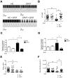
Contractile properties of CPVT human induced pluripotent stem cell-derived embryoid bodies (hiPSC-EBs). (A) Scatter plots showing beat rates for control human embryonic stem cell hESC-EBs (hESC), healthy control-EBs (HC) and CPVT-EBs (C2.2, CPVT) at rest. (B) Contraction force for hESC-, HC- and CPVT-EBs at rest. (C) Ratio of beat rates under 1 µM ISO to beat rates at rest for HC and CPVT ± (indicated by the + or − symbols) 5 µM S107 (overnight). (D) Ratio of contraction forces under 1 μM isoproterenol (ISO) to contraction forces at rest for HC and CPVT ± (indicated by the + or − symbols) 5 μM S107 (overnight). The dotted line indicates the ratio threshold. The number of experiments varies from 6 to 22 for each scatter plot. Data are presented with mean ± SEM. * p < 0.05; ** p < 0.01.
Figure 3
Figure 3
Intracellular Ca2+ cycling and SR Ca2+ stores in CPVT hiPSC-CMs under stress. (A) Display of original line-scan images of Ca2+ transients and corresponding tracings of HC and CPVT C2.2 hiPSC-CMs treated with 1 μM ISO together with pacing at 1 Hz (20 V, 0.5 ms duration and 1 ms delay) ± 5 μM S107. Additional and aberrant Ca2+ release events are shown with the arrows. (B) Scatter plots showing maximal Ca2+-transient amplitude in HC (white dots) and CPVT (black dots) hiPSC-CMs under stress conditions ± (indicated by the + or − symbols) treated with 5 μM S107. (C,D) Bar graphs showing frequency of occurrence of aberrant Ca2+-transients (C) and diastolic SR leaky events (D) in HC and CPVT hiPSC-CMs under stress conditions ± (indicated by the + or − symbols) treated with 5 μM S107. (E,F) Rate of RyR2 Ca2+ release (dF/dtmax) and area under the curve (peak area) in HC and CPVT hiPSC-CMs under stress conditions ± (indicated by the + or − symbols) treated with 5 μM S107. The number of experiments varies from 20 to 46 for each scatter plot. Data are presented with mean ± SEM. * p < 0.05; ** p < 0.01.
Figure 4
Figure 4
Flecainide (FLEC) partially prevents the aberrant SR Ca2+ handling in CPVT hiPSC-CMs. (A) Display of original line-scan images of Ca2+ transients and corresponding tracings of CPVT (C1.1) hiPSC-CMs after application of ±5 μM flecainide followed by 1 μM ISO. (B) Scatter plots showing maximal Ca2+-transient amplitude in CPVT hiPSC-CMs under ISO (white dots) and flecainide + ISO (black dots). (C) Bar graphs showing frequency of occurrence of aberrant Ca2+-transients in CPVT hiPSC-CMs under ISO and flecainide + ISO. (D) Frequency of occurrence of diastolic SR leaky events in CPVT hiPSC-CMs under ISO and flecainide + ISO. (E) Rate of RyR2 Ca2+ release (dF/dtmax) in CPVT hiPSC-CMs under ISO and flecainide + ISO. (F) Area under the curve or peak area in CPVT hiPSC-CMs under ISO and flecainide + ISO. The number of experiments varies from 10 to 55 for each scatter plot. Data are presented with mean ± SEM. ** p < 0.01.
Figure 5
Figure 5
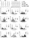
Action potential parameters from spontaneous electrical activity. (A) Example of spontaneous action potentials (AP) from HC (left) and CPVT (right) hiPSC-CM recorded using the patch-clamp technique on isolated cardiomyocytes. (BM) AP characteristics are studied and were compared between: HC (n = 26) and CPVT (n = 41) (BE), HC (n = 26) and HC + ISO (n = 20) (FI) and CPVT (n = 41) and CPVT + ISO (n = 29) (JM). For each comparison, the frequency distribution is shown with a scatter plot (with mean ± SEM) in inset to provide an accurate view of population distribution. The parameters studied were the spontaneous AP frequencies (B,F,J), the bazett’s corrected APD90 (C,G,K), the maximum depolarization speed (maximum dV/dt) (D,H,L) and the AP amplitude (E,I,M). * p < 0.05; ** p < 0.01, *** p < 0.001.
Figure 6
Figure 6
Abnormal electrical activity is elicited by ISO and normalized with flecainide and S107 treatments. (AC) Examples of current clamp raw traces showing APs recorded from CPVT hiPSC-CM after treatment with ISO (1 μM) (A), flecainide (5 μM) + ISO (B) and S017 + ISO (5 μM) (C). (DF) HC and CPVT abnormal electrical activity is studied with and without ISO (HC: n = 26, HC + ISO: n = 20, CPVT: n = 41, CPVT + ISO: n = 29). Percentages of variations are indicated in comparison with HC. (GI) The effect of flecainide (n = 12) and S107 (n = 16) pre-treatment (indicated by the + in contrast to -) on CPVT hiPSC-CM is also studied. Abnormal electrical activity is evaluated through the measure of the number of trace with aberrant events (D,G), the measure of abnormal event in each recording showing at least 1 abnormal event (E,H) and their frequency in Hz (F,I). * p < 0.05.
Figure 7
Figure 7
PKA-phosphorylated CPVT RyR2-D3638A mutant channels are depleted in Calstabin2 which is prevented by S107 but not by flecainide. (A) Representative immunoblot bands of HC and CPVT lysates (c2.2) at rest and under stress (1 μM ISO application) for total RyR2, PKA-phosphorylated RyR2 at S2809 site, spinophilin, PP2A and Calstabin2. In stress conditions, CPVT cells were also either pretreated with 5 μM flecainide (FLEC.), or 5 μM metoprolol (METO) or 5 μM S107 (BE) Bar graphs showing data normalization from immunoblots for the relative RyR2 PKA phosphorylation, spinophilin, PP2A and Calstabin2 bound to RyR2 in HC and CPVT at rest and under stress and in presence of flecainide, METO and S107. For each condition, n = 3 to 8 independent experiments. * p < 0.05; ** p < 0.01.
Figure 7
Figure 7
PKA-phosphorylated CPVT RyR2-D3638A mutant channels are depleted in Calstabin2 which is prevented by S107 but not by flecainide. (A) Representative immunoblot bands of HC and CPVT lysates (c2.2) at rest and under stress (1 μM ISO application) for total RyR2, PKA-phosphorylated RyR2 at S2809 site, spinophilin, PP2A and Calstabin2. In stress conditions, CPVT cells were also either pretreated with 5 μM flecainide (FLEC.), or 5 μM metoprolol (METO) or 5 μM S107 (BE) Bar graphs showing data normalization from immunoblots for the relative RyR2 PKA phosphorylation, spinophilin, PP2A and Calstabin2 bound to RyR2 in HC and CPVT at rest and under stress and in presence of flecainide, METO and S107. For each condition, n = 3 to 8 independent experiments. * p < 0.05; ** p < 0.01.

References

    1. Napolitano C., Bloise R., Memmi M., Priori S.G. Clinical utility gene card for: Catecholaminergic polymorphic ventricular tachycardia (CPVT) Eur. J. Hum. Genet. 2014;22 doi: 10.1038/ejhg.2013.55. - DOI - PMC - PubMed
    1. Sayed N., Liu C., Wu J.C. Translation of Human-Induced Pluripotent Stem Cells: From Clinical Trial in a Dish to Precision Medicine. J. Am. Coll. Cardiol. 2016;67:2161–2176. doi: 10.1016/j.jacc.2016.01.083. - DOI - PMC - PubMed
    1. Liang P., Sallam K., Wu H., Li Y., Itzhaki I., Garg P., Zhang Y., Termglichan V., Lan F., Gu M., et al. Patient-Specific and Genome-Edited Induced Pluripotent Stem Cell–Derived Cardiomyocytes Elucidate Single-Cell Phenotype of Brugada Syndrome. J. Am. Coll. Cardiol. 2016;68:2086–2096. doi: 10.1016/j.jacc.2016.07.779. - DOI - PMC - PubMed
    1. Peng W., Shen H., Wu J., Guo W., Pan X., Wang R., Chen S.R., Yan N. Structural basis for the gating mechanism of the type 2 ryanodine receptor RyR2. Science. 2016;354:aah5324. doi: 10.1126/science.aah5324. - DOI - PubMed
    1. Moreau A., Mercier A., Theriault O., Boutjdir M., Burger B., Keller D.I., Chahine M. Biophysical, Molecular, and Pharmacological Characterization of Voltage-Dependent Sodium Channels from Induced Pluripotent Stem Cell-Derived Cardiomyocytes. Can. J. Cardiol. 2017;33:269–278. doi: 10.1016/j.cjca.2016.10.001. - DOI - PubMed