. 2018 Nov 9;15(1):43.
doi: 10.1186/s12989-018-0280-2.

Group II innate lymphoid cells and microvascular dysfunction from pulmonary titanium dioxide nanoparticle exposure

Affiliations

Group II innate lymphoid cells and microvascular dysfunction from pulmonary titanium dioxide nanoparticle exposure

Alaeddin Bashir Abukabda et al. Part Fibre Toxicol. .

Abstract

Background: The cardiovascular effects of pulmonary exposure to engineered nanomaterials (ENM) are poorly understood, and the reproductive consequences are even less understood. Inflammation remains the most frequently explored mechanism of ENM toxicity. However, the key mediators and steps between lung exposure and uterine health remain to be fully defined. The purpose of this study was to determine the uterine inflammatory and vascular effects of pulmonary exposure to titanium dioxide nanoparticles (nano-TiO2). We hypothesized that pulmonary nano-TiO2 exposure initiates a Th2 inflammatory response mediated by Group II innate lymphoid cells (ILC2), which may be associated with an impairment in uterine microvascular reactivity.

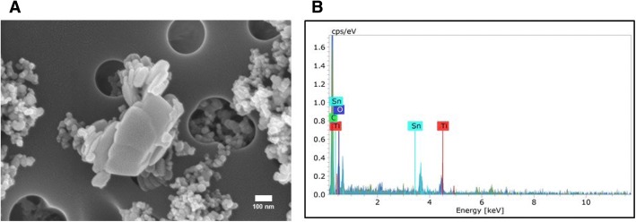
Methods: Female, virgin, Sprague-Dawley rats (8-12 weeks) were exposed to 100 μg of nano-TiO2 via intratracheal instillation 24 h prior to microvascular assessments. Serial blood samples were obtained at 0, 1, 2 and 4 h post-exposure for multiplex cytokine analysis. ILC2 numbers in the lungs were determined. ILC2s were isolated and phosphorylated nuclear factor kappa-light-chain-enhancer of activated B cells (NF-ĸB) levels were measured. Pressure myography was used to assess vascular reactivity of isolated radial arterioles.

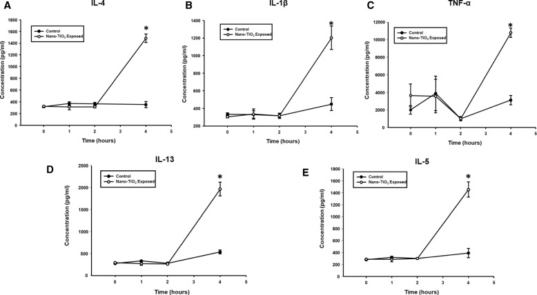
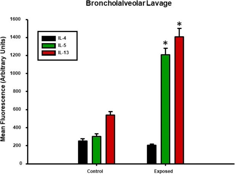
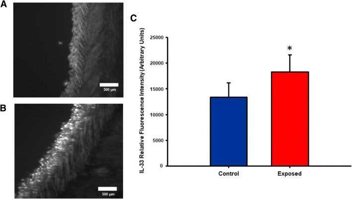
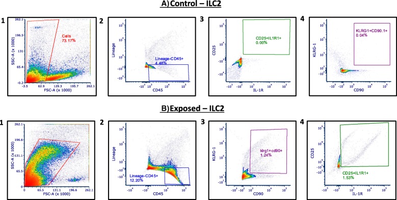
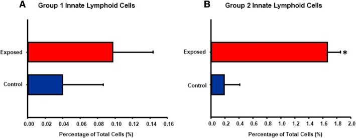
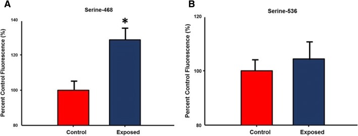
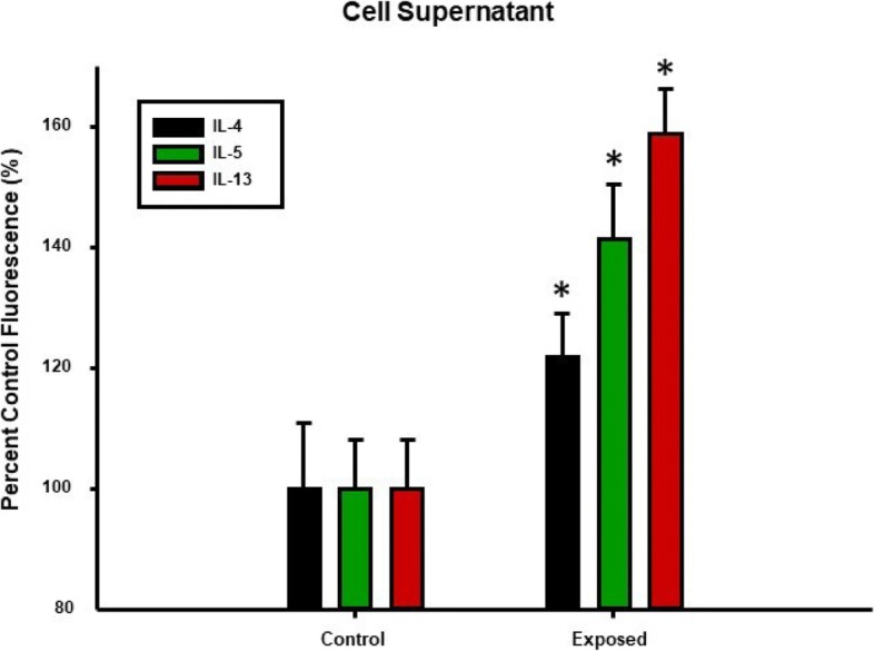
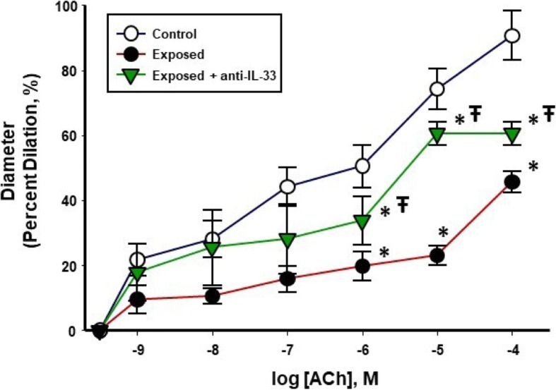
Results: Pulmonary nano-TiO2 exposure was associated with an increase in IL-1ß, 4, 5 and 13 and TNF- α 4 h post-exposure, indicative of an innate Th2 inflammatory response. ILC2 numbers were significantly increased in lungs from exposed animals (1.66 ± 0.19%) compared to controls (0.19 ± 0.22%). Phosphorylation of the transactivation domain (Ser-468) of NF-κB in isolated ILC2 and IL-33 in lung epithelial cells were significantly increased (126.8 ± 4.3% and 137 ± 11% of controls respectively) by nano-TiO2 exposure. Lastly, radial endothelium-dependent arteriolar reactivity was significantly impaired (27 ± 12%), while endothelium-independent dilation (7 ± 14%) and α-adrenergic sensitivity (8 ± 2%) were not altered compared to control levels. Treatment with an anti- IL-33 antibody (1 mg/kg) 30 min prior to nano-TiO2 exposure resulted in a significant improvement in endothelium-dependent dilation and a decreased level of IL-33 in both plasma and bronchoalveolar lavage fluid.

Conclusions: These results provide evidence that the uterine microvascular dysfunction that follows pulmonary ENM exposure may be initiated via activation of lung-resident ILC2 and subsequent systemic Th2-dependent inflammation.

Keywords: Engineered nanomaterials; Inflammation; Innate lymphoid cells; Microcirculation; Titanium dioxide nanoparticles.


Conflict of interest statement

Ethics approval and consent to participate

Not applicable.

Competing interests

The authors declare that they have no competing interests.

Publisher’s Note

Springer Nature remains neutral with regard to jurisdictional claims in published maps and institutional affiliations.

Figures

Fig. 1
Fig. 1
Characterization of Nano-TiO2. (a) SEM image and (b) Energy dispersive spectroscopy showing elemental composition of the nano-TiO2 suspension used in this study
Fig. 2
Fig. 2
Nano-TiO2 exposure increases T-Helper type II cytokines 4 h post-exposure. Multiplex cytokine analysis showing concentrations of IL-4 (a), IL-1β (b), TNF-α (c), IL-13 (d) and IL-5 (e). Serum samples were obtained at 0, 1, 2 and 4 h post-exposure via tail-vein puncture (N = 6). Statistics were analyzed with two-way ANOVA (P ≤ 0.05), * Sham control group vs. nano-TiO2 exposed groups
Fig. 3
Fig. 3
Nano-TiO2 exposure also increases T-Helper type II cytokines 4 h post-exposure in bronchoalveolar lavage fluid (BALF). Multiplex cytokine analysis showing concentrations of IL-4, IL-5, IL-13 in BALF from exposed and control animals 4 h post-exposure (N = 6). Statistics were analyzed with two-way ANOVA (P ≤ 0.05), * Sham control group vs. nano-TiO2 exposed groups
Fig. 4
Fig. 4
Nano-TiO2 exposure is associated with an increase in pulmonary interleukin-33 levels. Lung sections from control (a) and nano-TiO2 exposed (b) animals obtained 4 h post-exposure were stained for interleukin-33. Fluorescence was achieved by staining tracheal sections with an anti-IL-33-FITC conjugated antibody. Relative fluorescence is shown in (c) tagged antibody (N = 6–7). Statistics were analyzed with two-way ANOVA (P ≤ 0.05), * Sham control group vs. nano-TiO2 exposed groups
Fig. 5
Fig. 5
Pre-treatment of nano-TiO2 – exposed animals with an anti-IL-33 antibody lowers BALF and plasma IL-33. Interleukin-33 levels in (a) BALF and (b) plasma in control, nano-TiO2 exposed groups and in rats pre-treated with an anti-IL-33 antibody. Rats were pre-treated with an anti-IL-33 antibody (1 mg/kg) 30 min prior to exposure. Plasma and BALF IL-33 levels were measured 4 h post-exposure. Statistics were analyzed with two-way ANOVA (P ≤ 0.05). * Sham control group vs. nano-TiO2 exposed groups, Ŧ P < 0.05 Exposed + Anti-IL-33 Antibody vs Exposed
Fig. 6
Fig. 6
Flow cytometric analysis of lung-resident Group II innate lymphoid cells. Flow cytometry of lung tissue 4 h post-exposure for Group II Innate Lymphoid Cells in (a) control and (b) nano-TiO2 exposed animals (N = 6)
Fig. 7
Fig. 7
Nano-TiO2 exposure increases pulmonary ILC2 but not ILC1. Quantification of percentage of total pulmonary cells of (a) ILC1 and (b) ILC2 in control and nano-TiO2 exposed animals (N = 6). Statistics were analyzed with two-way ANOVA (P ≤ 0.05), * Sham control group vs. nano-TiO2 exposed groups
Fig. 8
Fig. 8
Nano-TiO2 exposure increases NF-ĸB phosphorylation at Serine-468 in isolated ILC2. Quantification of NF-ĸB phosphorylation at (a) serine-468 and (b) serine 536 in isolated ILC2 from isolated. ILC2 from control and nano-TiO2 exposed animals (N = 6). Statistics were analyzed with two-way ANOVA (P ≤ 0.05), * Sham control group vs. nano-TiO2 exposed groups
Fig. 9
Fig. 9
Th2 cytokine secretion in isolated ILC2 is increased by nano-TiO2 exposure. Multiplex cytokine analysis showing concentrations of IL-4, IL-5, IL-13 supernatant from isolated ILC2 cultured overnight from control and nano-TiO2 exposed animals (N = 6). Statistics were analyzed with two-way ANOVA (P ≤ 0.05), * Sham control group vs. nano-TiO2 exposed groups
Fig. 10
Fig. 10
Endothelium-dependent dilation of radial arterioles is blunted by nano-TiO2 exposure and improved by pre-treatment with an anti-IL-33 antibody. Endothelium-dependent dilation of uterine radial arterioles from control, exposed and anti-IL-33 antibody treated animals was determined using pressure myography (n = 12–18). All vascular assessments were performed 24 h post-exposure. Statistics were analyzed with two-way ANOVA (P ≤ 0.05). * Sham control group vs. nano-TiO2 exposed groups, Ŧ P < 0.05 Exposed + Anti-IL-33 Antibody vs Exposed

References

    1. Jackson P, et al. Prenatal exposure to carbon black (printex 90): effects on sexual development and neurofunction. Basic Clin Pharmacol Toxicol. 2011;109(6):434–437. doi: 10.1111/j.1742-7843.2011.00745.x. - DOI - PubMed
    1. Johansson HKL, et al. Airway exposure to multi-walled carbon nanotubes disrupts the female reproductive cycle without affecting pregnancy outcomes in mice. Part Fibre Toxicol. 2017;14(1):17. doi: 10.1186/s12989-017-0197-1. - DOI - PMC - PubMed
    1. Stapleton PA, et al. Maternal engineered nanomaterial exposure and fetal microvascular function: does the barker hypothesis apply? Am J Obstet Gynecol. 2013;209(3):227 e1–227 11. doi: 10.1016/j.ajog.2013.04.036. - DOI - PMC - PubMed
    1. Stapleton PA, et al. Maternal engineered nanomaterial inhalation during gestation alters the fetal transcriptome. Part Fibre Toxicol. 2018;15(1):3. doi: 10.1186/s12989-017-0239-8. - DOI - PMC - PubMed
    1. Engler-Chiurazzi EB, et al. Impacts of prenatal nanomaterial exposure on male adult Sprague-Dawley rat behavior and cognition. J Toxicol Environ Health A. 2016;79(11):447–452. doi: 10.1080/15287394.2016.1164101. - DOI - PMC - PubMed

Publication types

MeSH terms

LinkOut - more resources