

Case Reports
doi: 10.1016/j.jpeds.2018.10.018.
Epub 2018 Nov 7.
Neonatal Lung Disease Associated with TBX4 Mutations
Affiliations
- PMID: 30413314
- PMCID: PMC6389379
- DOI: 10.1016/j.jpeds.2018.10.018
Item in Clipboard
Case Reports
Neonatal Lung Disease Associated with TBX4 Mutations
J Pediatr.
2019 Mar.
Abstract
Variable lung disease was documented in 2 infants with heterozygous TBX4 mutations; their clinical presentations, pathology, and outcomes were distinct. These findings demonstrate that TBX4 gene mutations are associated with neonatal respiratory failure and highlight the wide spectrum of clinicopathological outcomes that have implications for patient diagnosis and management.
Keywords: ABCA3; T-box transcription factor; TBX2; TBX4; congenital alveolar dysplasia; congenital anomaly; lung development; pulmonary hypoplasia.
Copyright © 2018 Elsevier Inc. All rights reserved.
Figures
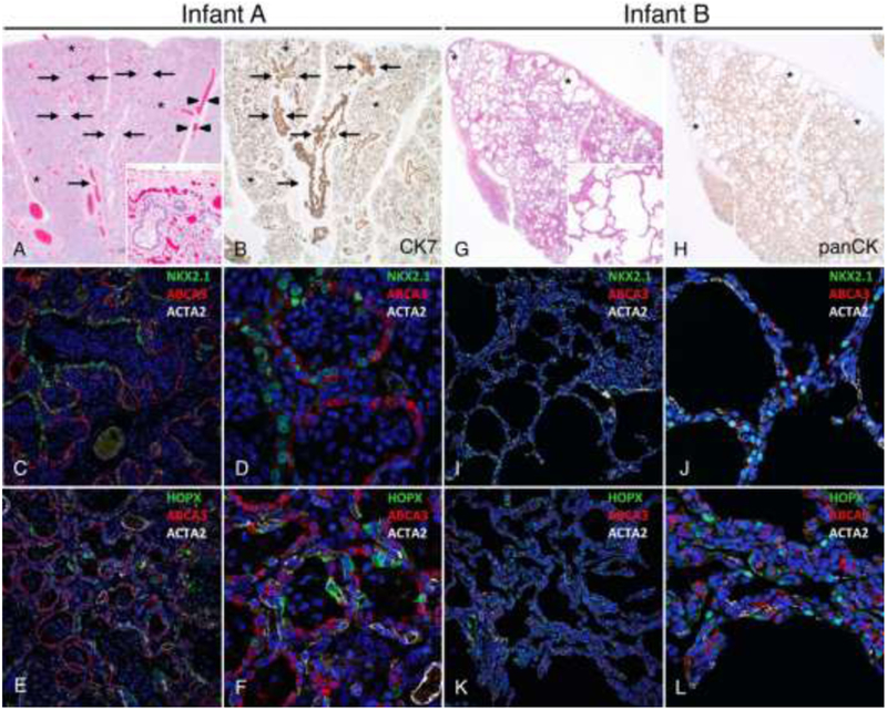
Lungs from Infant A (A-F) showing congenital alveolar dysplasia. H&E stained section (A) shows normally developed branching bronchi and bronchioles accompanied by arteries (arrows), veins appropriately located in the interlobular septa (arrowheads), and malformed lobules comprised of variable numbers of simplified saccular airspaces (*) with bronchioles focally abutting the pleura (inset). Epithelial cell immunostaining with cytokeratin 7 (B) highlights branching conducting airways (arrows) and variable numbers of simplified saccules comprising the malformed lobules (*). Confocal immunofluorescence low (C,E) and higher power images (D,F) show aberrant lack of co-expression of the alveolar type II cell (AT2) markers NKX2.1 and ABCA3 in the atypical epithelial cells lining distal saccular airspaces (C-D) and absence of nuclear staining for the alveolar type I cell marker (AT1), HOPX (E-F). Staining for smooth muscle actin (ACTA2) is also shown. Lungs from Infant B (G-L) showing alveolar growth abnormality. H&E stained section shows deficient alveolarization with variably enlarged and simplified alveoli (inset) and subpleural cystically dilated airspaces (*). Epithelial cell immunostaining with pancytokeratin (H) highlights the focally enlarged and simplified alveolar spaces that predominant in subpleural regions (*). In contrast to Infant A, confocal immunofluorescence low (I,K) and higher power images (J, L) of the lung biopsy from Infant B show co-expression of NKX2.1 and ABCA3 as seen in normal AT2 cell differentiation (I-J) and nuclear HOPX expression indicative of AT1 cell differentiation (K-L). Original magnification: 20x (A-B, G-H); 200x (A inset, G inset); 20X objective with pixel size of 0.63 μm (C, E, I, K); 60X objective with pixel size of 0.21 μm (D, F, J, L).
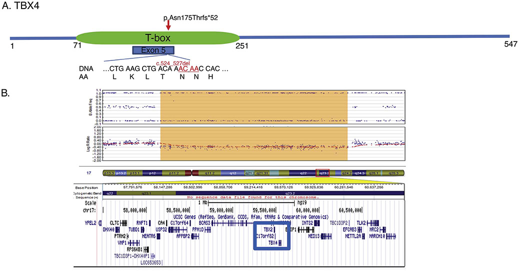
A) Whole exome sequencing performed on Infant A identified a de novo four base pair deletion in exon 5 (c.524_527del) located within the T-box, the critical DNA binding domain of the resultant protein. B) Microarray analysis performed on Infant B identified a 2.2Mb deletion in the long arm of chromosome 17 (17q23.1-17q23.2; 58113570_60325222) encompassing the TBX2 and TBX4 genes.
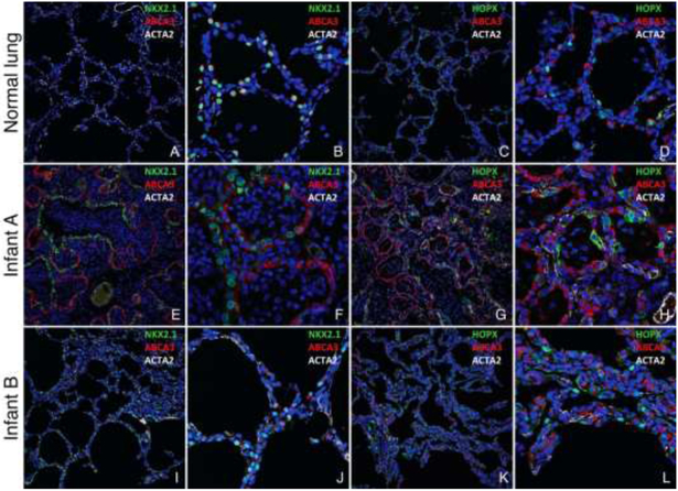
Confocal immunofluorescence images of lungs from a 4 month old donor control lung (A-D), Infant A (E-H) and Infant B (I-L). Nuclear NKX2.1 and cytoplasmic ABCA3 are co-expressed in AT2 cells (A-B), and nuclear HOPX is expressed in AT1 cells (C-D) in the normal control lung. In contrast, the distal saccules comprising the lobules in Infant A are lined by atypical epithelial cells with a discrepancy in NKX2.1 and ABCA3 expression (E-F), and lack of nuclear HOPX expression (G-H). Enlarged and simplified alveoli in Infant B are lined by AT2 cells co-expressing NKX2.1 and ABCA3 (I-J) and AT1 cells expressing nuclear HOPX, Original magnification: 20X objective with pixel size of 0.63 μm (A, C, E, G, I, K), 60X objective with pixel size of 0.21 μm (B, D, F, H, J, L).
References
-
- Chow CW, Massie J, Ng J, Mills J, Baker M. Acinar dysplasia of the lungs: variation in the extent of involvement and clinical features. Pathology. 2013;45:38–43. - PubMed
-
- Langston C, Dishop MK. Diffuse lung disease in infancy: a proposed classification applied to 259 diagnostic biopsies. Pediatr Dev Pathol. 2009;12:421–37. - PubMed
-
- Barnett CP, Nataren NJ, Klingler-Hoffmann M, Schwarz Q, Chong CE, Lee YK, et al. Ectrodactyly and Lethal Pulmonary Acinar Dysplasia Associated with Homozygous FGFR2 Mutations Identified by Exome Sequencing. Human mutation. 2016;37:955–63. - PubMed
Publication types
MeSH terms
Substances
Grants and funding
LinkOut - more resources
Full Text Sources
Medical
