. 2019 Jan 9;85(2):e02129-18.
doi: 10.1128/AEM.02129-18. Print 2019 Jan 15.

Manganese Oxide Biomineralization Provides Protection against Nitrite Toxicity in a Cell-Density-Dependent Manner

Affiliations

Manganese Oxide Biomineralization Provides Protection against Nitrite Toxicity in a Cell-Density-Dependent Manner

Christian Zerfaß et al. Appl Environ Microbiol. .

Abstract

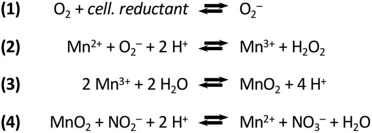
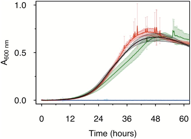
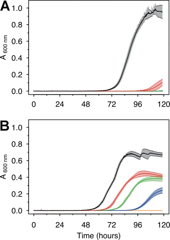
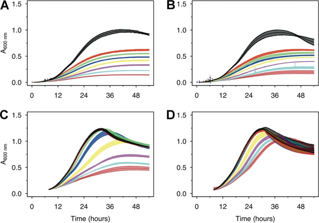
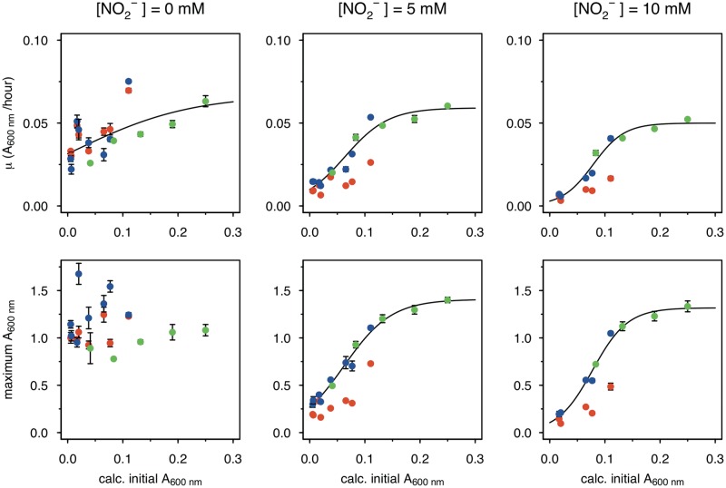
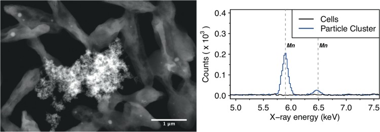
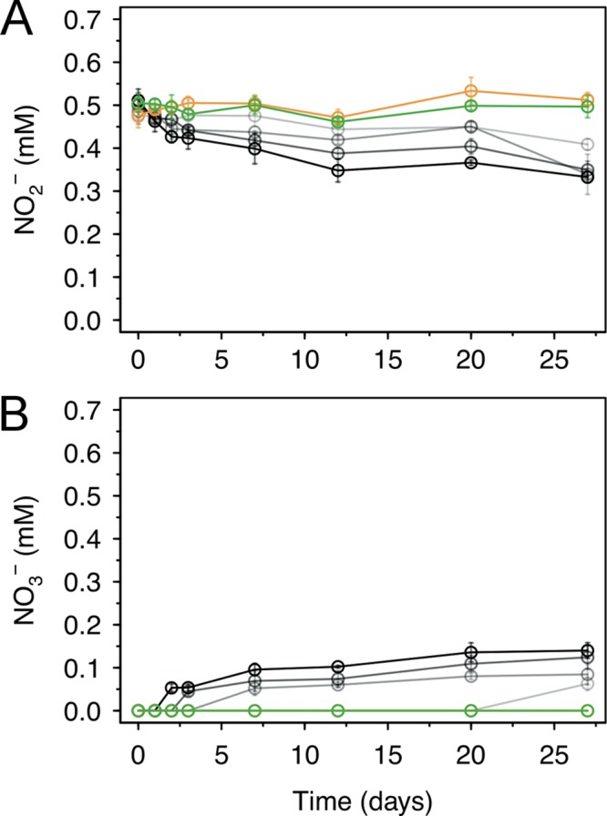
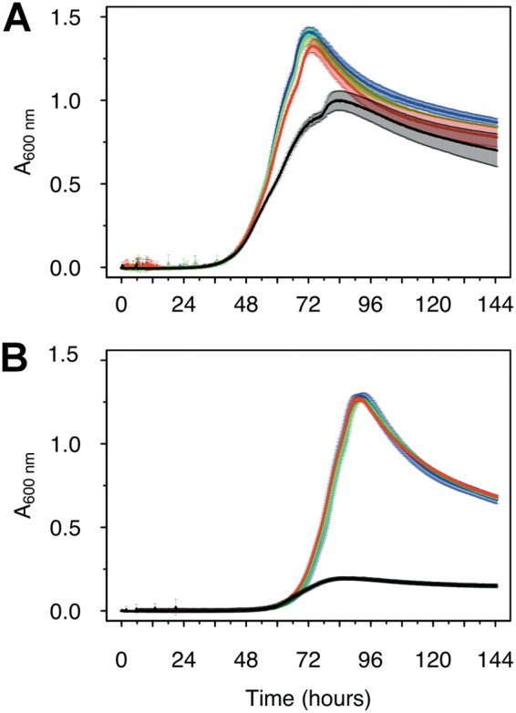
Manganese biomineralization is a widespread process among bacteria and fungi. To date, there is no conclusive experimental evidence for how and if this process impacts microbial fitness in the environment. Here, we show how a model organism for manganese oxidation is growth inhibited by nitrite, and that this inhibition is mitigated in the presence of manganese. We show that such manganese-mediated mitigation of nitrite inhibition is dependent on the culture inoculum size, and that manganese oxide (MnOX) forms granular precipitates in the culture, rather than sheaths around individual cells. We provide evidence that MnOX protection involves both its ability to catalyze nitrite oxidation into (nontoxic) nitrate under physiological conditions and its potential role in influencing processes involving reactive oxygen species (ROS). Taken together, these results demonstrate improved microbial fitness through MnOX deposition in an ecological setting, i.e., mitigation of nitrite toxicity, and point to a key role of MnOX in handling stresses arising from ROS.IMPORTANCE We present here a direct fitness benefit (i.e., growth advantage) for manganese oxide biomineralization activity in Roseobacter sp. strain AzwK-3b, a model organism used to study this process. We find that strain AzwK-3b in a laboratory culture experiment is growth inhibited by nitrite in manganese-free cultures, while the inhibition is considerably relieved by manganese supplementation and manganese oxide (MnOX) formation. We show that biogenic MnOX interacts directly with nitrite and possibly with reactive oxygen species and find that its beneficial effects are established through formation of dispersed MnOX granules in a manner dependent on the population size. These experiments raise the possibility that manganese biomineralization could confer protection against nitrite toxicity to a population of cells. They open up new avenues of interrogating this process in other species and provide possible routes to their biotechnological applications, including in metal recovery, biomaterials production, and synthetic community engineering.

Keywords: Roseobacter; biomineralization; metal recovery; microbial ecology; reactive oxygen species; respiration.


Figures

FIG 1
FIG 1
Biological oxidation of manganese via superoxide and nitrite oxidation by the product manganese oxide. These reactions are taken from references (manganese oxidation) and (nitrite oxidation). Note that only representative reactions are presented. For instance, the text refers to a mixed oxide (MnOX), while this reaction scheme simplifies to MnO2. The cellular reductant (cell. reductant) which serves as electron donor for superoxide production is not unambiguously identified.
FIG 2
FIG 2
Effect of MnII on the growth of Roseobacter sp. AzwK-3b in the defined growth medium (Table 1). The concentrations of manganese were 0 μM (black), 200 μM (red), and 500 μM (dark green), with no growth (zero line) in the respective noninoculated controls (blue, magenta, and light blue). Cultures were grown in a 96-well plate (200-μl culture), with shaking, and absorbance measurements were taken every 10 min (see Materials and Methods).
FIG 3
FIG 3
Growth of Roseobacter sp. AzwK-3b cultures in the defined growth medium supplemented with sodium nitrite. Media were prepared without (A) or with (B) 200 μM manganese chloride (MnIICl2). Nitrite concentrations were 0 mM (black), 0.25 mM (red), 0.5 mM (green), 1 mM (dark blue), and 2.5 mM (light blue). All conditions were tested in triplicate, and the growth curves represent averages and their standard deviations (see Materials and Methods).
FIG 4
FIG 4
Larger AzwK-3b inocula are less inhibited by nitrite. A preculture without manganese or nitrite was grown and sampled in the exponential growth phase (Fig. S4) to prepare inocula from a very early time point in the exponential phase (IT1, panels A and B) and from a later time point (IT2, panels C and D; both sampled in the first third of the exponential phase). These inocula were diluted 1:1 with fresh medium and tested for growth at different nitrite concentrations (see below for color code) without (A, C) or with (B, D) 200 μM MnIICl2 supplement. The nitrite concentrations were as follows: black, control/no nitrite; red, 0.25 mM nitrite; green, 0.5 mM nitrite; blue, 1 mM nitrite; yellow, 2 mM nitrite; magenta, 5 mM nitrite; light blue, 7.5 mM nitrite; and dark red, 10 mM nitrite. Growth curves show the averages and standard deviations over a triplicate analysis (see Materials and Methods).
FIG 5
FIG 5
Inoculum size effect on MnOX-mediated mitigation of nitrite inhibition. Data from different AzwK-3b growth experiments of similar type (large inocula; see Materials and Methods) were analyzed for the maximum A600 (bottom row) and growth rate (top row) by fitting the growth curves. Each condition was done in three technical replicates (note that error bars are not visible in some cases due to only small differences). Nitrite concentrations of the main cultures are indicated as headings of the figure columns. The x axes show the calculated A600 values of the initial cultures after diluting them 1:1 from the precultures, while the y axes show the maximum A600 and maximum growth rate values as calculated with the Gompertz model (91, 92) (see Materials and Methods). The colors represent different conditions, as follows: red, neither preculture nor main culture contained manganese; blue, preculture without and main culture with manganese; and green, both preculture and main culture with manganese. The black curve is a sigmoidal fit (logistic model) from the R package Grofit (91), for the results of the combined blue and green data set, where the nitrite-exposed main cultures all contained manganese.
FIG 6
FIG 6
Scanning transmission electron micrograph (left figure, high-angle annular dark field) of (granular) manganese-containing precipitate (center) surrounded by AzwK-3b cells, and associated energy-dispersive X-ray spectroscopic analysis (right figure) in this location. Only the energy range containing the manganese-specific X-ray energies at 5.90 keV (KαI) and 6.49 keV (KβI) is shown, and the manganese transitions are indicated by vertical gray dashed lines.
FIG 7
FIG 7
Oxidation of nitrite by biogenic manganese oxide (MnOX) produced in cell-free culture supernatant of AzwK-3b. The figure shows the concentrations over time of nitrite (A) and nitrate (B), as determined by ion chromatography (note that concentrations were corrected for the IC peak from chloride, to account for evaporation during the experiment). As controls, samples without MnOX (green) or with MnO2 powder (orange) were included in the experiment (see Materials and Methods). The samples with AzwK-3b cell-free manganese oxide contained (from gray to black) 0.2, 0.5, 1, and 2 mM manganese oxide equivalent (see Materials and Methods).
FIG 8
FIG 8
Reductive power (NADH) mitigates the growth inhibitory effects of nitrite in AzwK-3b. Cultures (preculture and main culture without manganese) were grown in the absence (A) and presence (B) of 5 mM nitrite and supplement of 0, 50, 100, and 200 μM NADH (black, red, green, and blue) at the start of the culture.

References

    1. Lowenstam H. 1981. Minerals formed by organisms. Science 211:1126–1131. doi:10.1126/science.7008198. - DOI - PubMed
    1. Hansel CM. 2017. Manganese in marine microbiology, p 37–83. In Poole RK. (ed), Advances in microbial physiology: microbiology of metal ions. Academic Press, Oxford, UK. - PubMed
    1. Nealson KH. 2006. The manganese-oxidizing bacteria. Prokaryotes 5:222–231.
    1. Ghiorse WC. 1984. Biology of iron-and manganese-depositing bacteria. Annu Rev Microbiol 38:515–550. doi:10.1146/annurev.mi.38.100184.002503. - DOI - PubMed
    1. Spiro TG, Bargar JR, Sposito G, Tebo BM. 2010. Bacteriogenic manganese oxides. Acc Chem Res 43:2–9. doi:10.1021/ar800232a. - DOI - PubMed

Publication types

LinkOut - more resources