Review
. 2018 Nov;75(11):481-494.
doi: 10.1002/cm.21504. Epub 2018 Dec 7.

Regulating a key mitotic regulator, polo-like kinase 1 (PLK1)

Affiliations
Review

Regulating a key mitotic regulator, polo-like kinase 1 (PLK1)

Erica G Colicino et al. Cytoskeleton (Hoboken). 2018 Nov.

Abstract

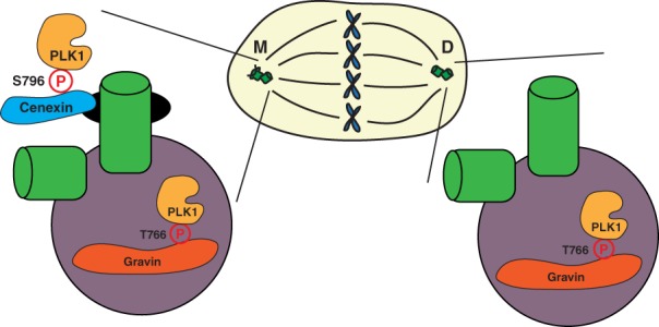
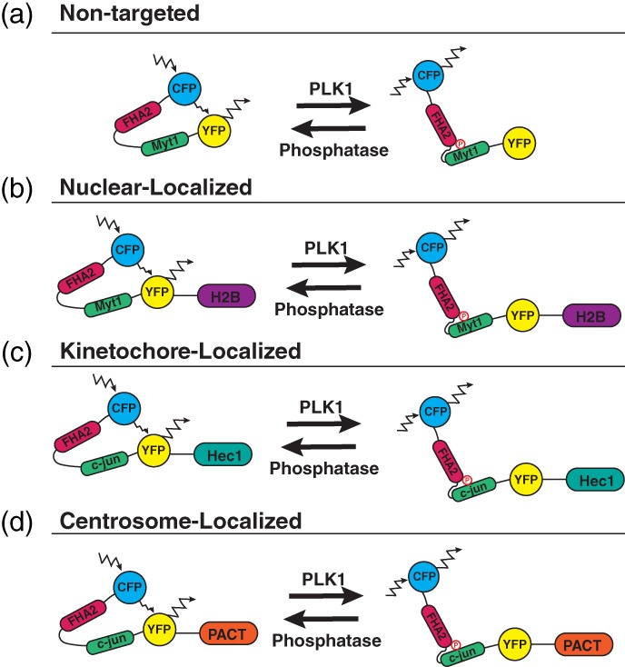
During cell division, duplicated genetic material is separated into two distinct daughter cells. This process is essential for initial tissue formation during development and to maintain tissue integrity throughout an organism's lifetime. To ensure the efficacy and efficiency of this process, the cell employs a variety of regulatory and signaling proteins that function as mitotic regulators and checkpoint proteins. One vital mitotic regulator is polo-like kinase 1 (PLK1), a highly conserved member of the polo-like kinase family. Unique from its paralogues, it functions specifically during mitosis as a regulator of cell division. PLK1 is spatially and temporally enriched at three distinct subcellular locales; the mitotic centrosomes, kinetochores, and the cytokinetic midbody. These localization patterns allow PLK1 to phosphorylate specific downstream targets to regulate mitosis. In this review, we will explore how polo-like kinases were originally discovered and diverged into the five paralogues (PLK1-5) in mammals. We will then focus specifically on the most conserved, PLK1, where we will discuss what is known about how its activity is modulated, its role during the cell cycle, and new, innovative tools that have been developed to examine its function and interactions in cells.

Keywords: FRET; biosensor; cell division; centrosome; chemical genetics; kinetochore; midbody; mitosis; polo-like kinase 1; scaffolds.


Figures

Figure 1
Figure 1
There are five mammalian polo‐like kinase paralogues. The five mammalian paralogues contain an N‐terminal catalytic kinase domain (orange) and a C‐terminal polo‐box domain (PBD, purple). The PLK4 cryptic polo‐box domain (CPB) is shown in green. Adapted from (De Cárcer, Manning, et al., 2011)
Figure 2
Figure 2
PLK1 subcellular distribution and function during metaphase in mammalian cells. (a) PLK1 (orange) localizes during M‐phase to the mitotic centrosomes, kinetochores, and cytokinetic midbody (magenta) to ensure mitotic progression, microtubule attachments, and anaphase onset, as well as proper cytokinesis and abscission. Gradient below (orange) represents relative PLK1 activity changes between prometaphase/metaphase and cytokinesis. (b) Table outlining PLK1 localization patterns with corresponding functions and known binding scaffolds during M‐phase
Figure 3
Figure 3
PLK1 distribution throughout mitosis. Structured illumination microscopy (SIM) volumetric projection micrographs of RPE cells showing localization of PLK1 (green) from interphase through cytokinesis. Kinetochore marker: CREST (red). Unpublished SIM micrographs from Dr. Heidi Hehnly's lab, performed by Erica Colicino
Figure 4
Figure 4
Model of PLK1 chemical genetics. The catalytic domain of PLK1 can be inhibited by treatment with an ATP analogue, such as the drug BI2536. By mutating and enlarging this catalytic domain, PLK1 can be inhibited in cells expressing this mutant using a purine analogue. Adapted from Burkard et al. (2007)
Figure 5
Figure 5
PLK1 scaffolding at the mitotic centrosomes. A model depicting the localization of PLK1 (gold) to the mother centriole appendages (black) and pericentriolar matrix (PCM, purple) through its binding scaffolds, cenexin (blue) and Gravin (red). PLK1 has been shown to bind cenexin, a known mother centriole appendage protein, at its phosphorylated S796 site (Soung et al., 2009). PLK1 has also been shown to bind Gravin, a known PCM component, at its phosphorylated T766 site (Canton et al., 2012). These scaffolds subsequently sequester PLK1 at its subcentrosomal locales, regulating its activity during mitosis
Figure 6
Figure 6
PLK1 FRET biosensors. (a) A nontargeted PLK1 FRET biosensor containing a CFP monomer (blue), FHA2 phospho‐binding domain (magenta), Myt1 PLK1‐substrate sequence (green), and YFP monomer (yellow). When active‐PLK1 is present, it phosphorylates the Myt1 sequence (T495), causing a conformational change in the biosensor, decreasing FRET. When no active‐PLK1 is present, the biosensor is in a relaxed state, allowing for FRET (Macůrek et al., 2008). (b) A nuclear‐localized PLK1 FRET‐biosensor fused to H2B (purple) (Bruinsma, Raaijmakers, & Medema, 2012). (c) A kinetochore‐localized PLK1 FRET‐biosensor containing a c‐jun PLK1‐susbtrate sequence and fused to Hec1 (cyan). The c‐jun substrate sequence was mutated (S17T), allowing for PLK1‐specific phosphorylation of the biosensor (Liu et al., 2012). (d) A centrosome‐localized PLK1 FRET‐biosensor containing a c‐jun PLK1‐substrate sequence and fused to the pericentrin AKAP centrosomal‐targeting (PACT) domain (orange) (Colicino et al., 2018)

References

    1. Agromayor, M. , Carlton, J. G. , Phelan, J. P. , Matthews, D. R. , Carlin, L. M. , Ameer‐Beg, S. , … Martin‐Serrano, J. (2009). Essential role of hIST1 in cytokinesis. Molecular Biology of the Cell, 20, 1374–1387. 10.1091/mbc.E08 - DOI - PMC - PubMed
    1. Andrysik, Z. , Bernstein, W. Z. , Deng, L. , Myer, D. L. , Li, Y.‐Q. , Tischfield, J. A. , … Mustapha Bahassi, E. (2010). The novel mouse polo‐like kinase 5 responds to DNA damage and localizes in the nucleolus. Nucleic Acids Research, 38, 2931–2943. - PMC - PubMed
    1. Awad, M. M. , Chu, Q. S. C. , Gandhi, L. , Stephenson, J. J. , Govindan, R. , Bradford, D. S. , … Socinski, M. A. (2017). An open‐label, phase II study of the polo‐like kinase‐1 (Plk‐1) inhibitor, BI 2536, in patients with relapsed small cell lung cancer (SCLC). Lung Cancer, 104, 126–130. 10.1016/j.lungcan.2016.12.019 - DOI - PubMed
    1. Bastos, R. N. , & Barr, F. A. (2010). Plk1 negatively regulates Cep55 recruitment to the midbody to ensure orderly abscission. Journal of Cell Biology, 191(4), 751–760. 10.1083/jcb.201008108 - DOI - PMC - PubMed
    1. Beck, J. , Maerki, S. , Posch, M. , Metzger, T. , Persaud, A. , Scheel, H. , … Sumara, I. (2013). Ubiquitylation‐dependent localization of PLK1 in mitosis. Nature Cell Biology, 15(4), 430–439. 10.1038/ncb2695 - DOI - PMC - PubMed

Publication types

Substances

LinkOut - more resources