Review
. 2018 Dec:76:101-115.
doi: 10.1016/j.ceca.2018.10.004. Epub 2018 Oct 30.

Role of STIM1/ORAI1-mediated store-operated Ca2+ entry in skeletal muscle physiology and disease

Affiliations
Review

Role of STIM1/ORAI1-mediated store-operated Ca2+ entry in skeletal muscle physiology and disease

Antonio Michelucci et al. Cell Calcium. 2018 Dec.

Abstract

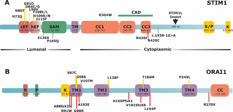
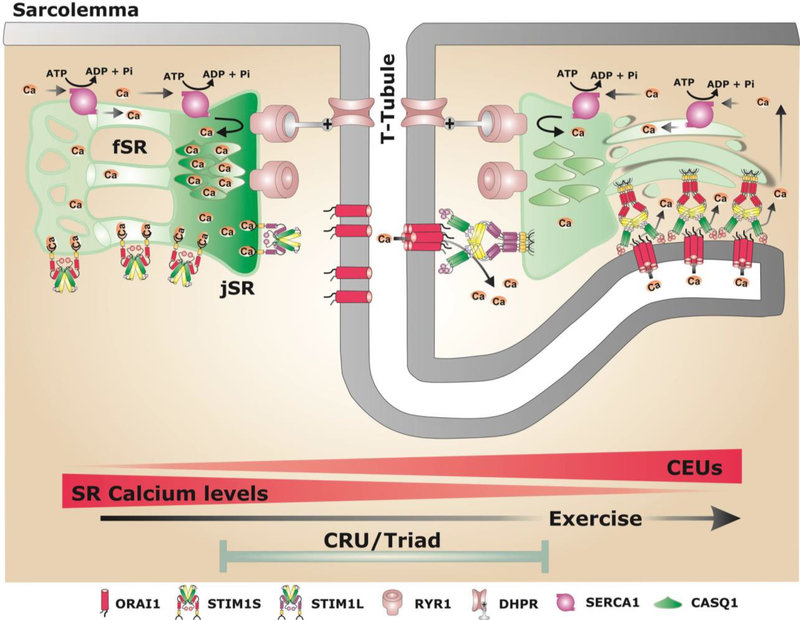
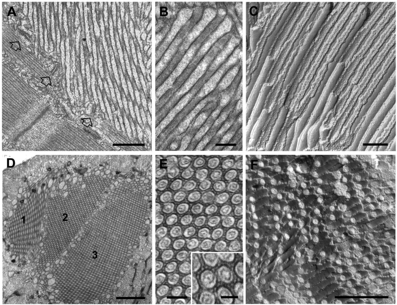
Store-operated Ca2+ entry (SOCE) is a Ca2+ entry mechanism activated by depletion of intracellular Ca2+ stores. In skeletal muscle, SOCE is mediated by an interaction between stromal-interacting molecule-1 (STIM1), the Ca2+ sensor of the sarcoplasmic reticulum, and ORAI1, the Ca2+-release-activated-Ca2+ (CRAC) channel located in the transverse tubule membrane. This review focuses on the molecular mechanisms and physiological role of SOCE in skeletal muscle, as well as how alterations in STIM1/ORAI1-mediated SOCE contribute to muscle disease. Recent evidence indicates that SOCE plays an important role in both muscle development/growth and fatigue. The importance of SOCE in muscle is further underscored by the discovery that loss- and gain-of-function mutations in STIM1 and ORAI1 result in an eclectic array of disorders with clinical myopathy as central defining component. Despite differences in clinical phenotype, all STIM1/ORAI1 gain-of-function mutations-linked myopathies are characterized by the abnormal accumulation of intracellular membranes, known as tubular aggregates. Finally, dysfunctional STIM1/ORAI1-mediated SOCE also contributes to the pathogenesis of muscular dystrophy, malignant hyperthermia, and sarcopenia. The picture to emerge is that tight regulation of STIM1/ORAI1-dependent Ca2+ signaling is critical for optimal skeletal muscle development/function such that either aberrant increases or decreases in SOCE activity result in muscle dysfunction.

Keywords: Ca(2+) signaling; Ca(2+)-release-activated-Ca(2+) (CRAC); Muscle fatigue; Tubular aggregate myopathy (TAM).


Figures

Figure 1.
Figure 1.. Schematic representation of STIM1 and ORAI1 proteins and location of associated disease mutations.
A) STIM1 protein structure. cEF, canonical EF-hand; hEF, hidden EF-hand; SAM, sterile α-motif; TM, transmembrane; CC, coiled-coil region; S/P serine-proline-rich domain; K, lysine-rich domain; CAD, channel activation domain. B) ORAI1 protein structure. R/P, arginine-proline-rich domain; TM, transmembrane domain; CC, coiled-coil domain. Upper, yellow lines indicate gain-of-function mutations. Lower, red lines indicate loss-of-function mutations.
Figure 2.
Figure 2.. Schematic model showing potential sites of STIM1/ORAI1 coupling in skeletal muscle under resting conditions and after exercise.
The SR of adult fibers is divided in two compartments: i) the junctional SR (jSR, or SR terminal cisternae) that contains CASQ1 and RYR1 and is closely associated with the T-tubule (TT) that contains DHPR, to form the triad (or CRU), the site of excitation-contraction (EC) coupling; ii) the free SR (fSR) that does not interact with TT and is localized throughout I band. Both jSR and fSR contain high levels of the sarco/endoplasmic Ca2+ ATPase-1 (SERCA1), a Ca2+ ATPase that pumps Ca-2+ ions Ca2+ released during EC coupling from the myoplasm back into the lumen of the SR. Two different splice variants are expressed in skeletal muscle: STIM1S and STIM1L. Under resting conditions (left side), ORAI1 is located within the TT system at the triad, while STIM1S and STIM1L are located in the fSR and jSR, respectively. After exercise (right side): i) the fSR and TT undergo a striking remodeling to form new junctions composed by multiple layers of flat parallel stacks of fSR cisternae and an extension of the TT from the triad into the I-band toward the Z-line [21]. These new junctions promote increased STIM1/ORAI1 colocalization at the I-band as TT elongation allows ORAI1 to interact with STIM1S proteins in the fSR. These new junctions are proposed to function as Calcium Entry Units (CEUs) [21].
Figure 3.
Figure 3.. Tubular aggregates (TAs) in extensor digitorum longus fibers from 2 year old male wild type mice.
A-C) EM images of longitudinal views of TAs in thin sections (A and B) and freeze fracture (C). Empty arrows in panel A point to jSR-TT junctions (or triads) located at the periphery of a TA. Tubes of TAs are typically filled with electron-dense material, most likely CASQ1 (B). The regular and straight shape of the tubules are emphasized in freeze fracture replicas, where tubules appear as long cylinders showing alternated views of the luminal and cytoplasmic leaflets (see [75] for additional detail). D-F) EM images of cross-sectional views of TAs in thin sections (D and E) and freeze fracture (F). Large TAs often result from the association of multiple TAs of smaller size (panel D: 1–3). In the core of each domain, tubes forming the TA display uniform diameters and appear ordered in a hexagonal pattern (E). Note the presence of small linkages that bridge the gap between membranes of adjacent tubules (E, inset). Bars: A, D and F: 0.5 μm; B and C: 0.2 μm; E: 0.1 μm (inset: 0.05 μm)

References

    1. Putney JW Jr., A model for receptor-regulated calcium entry, Cell Calcium, 7 (1986) 1–12. - PubMed
    1. Hoth M, Penner R, Depletion of intracellular calcium stores activates a calcium current in mast cells, Nature, 355 (1992) 353–356. - PubMed
    1. Roos J, DiGregorio PJ, Yeromin AV, Ohlsen K, Lioudyno M, Zhang S, Safrina O, Kozak JA, Wagner SL, Cahalan MD, Velicelebi G, Stauderman KA, STIM1, an essential and conserved component of store-operated Ca2+ channel function, J Cell Biol, 169 (2005) 435–445. - PMC - PubMed
    1. Feske S, Gwack Y, Prakriya M, Srikanth S, Puppel SH, Tanasa B, Hogan PG, Lewis RS, Daly M, Rao A, A mutation in Orai1 causes immune deficiency by abrogating CRAC channel function, Nature, 441 (2006) 179–185. - PubMed
    1. Prakriya M, Lewis RS, Store-Operated Calcium Channels, Physiol Rev, 95 (2015) 1383–1436. - PMC - PubMed

Publication types