. 2019 Mar:120:285-296.
doi: 10.1016/j.bone.2018.11.003. Epub 2018 Nov 7.

Deletion of nicotinic acetylcholine receptor alpha9 in mice resulted in altered bone structure

Affiliations

Deletion of nicotinic acetylcholine receptor alpha9 in mice resulted in altered bone structure

Lisa Baumann et al. Bone. 2019 Mar.

Abstract

Alterations in bone strength and structure were found in knockout (KO) mouse strains with deletion of several acetylcholine receptors. Interestingly, the expression of the nicotinic acetylcholine receptors (nAChR) subunit α10 was down-regulated in osteogenic differentiated mesenchymal stem cells of patients with osteoporosis whereas the expression of subunit α9 was not altered. Since nAChR subunits α9 and α10 are often combined in a functional receptor, we analyzed here the bone of adult female KO mice with single deletion of either nAChR alpha9 (α9KO) or alpha10 (α10KO). Biomechanical testing showed a significant decrease of bending stiffness and maximal breaking force in α9KO compared to their corresponding wild type mice. Furthermore, an increase in trabecular pattern factor (Tb.Pf) and structure model index (SMI) was detected by μCT in α9KO indicating reduced bone mass. On the mRNA level a decrease of Collagen 1α1 and Connexin-43 was measured by real-time RT-PCR in α9KO while no alteration of osteoclast markers was detected in either mouse strain. Using electron microcopy we observed an increase in the number of osteocytes that showed signs of degeneration and cell death in the α9KO compared to their wild type mice, while α10KO showed no differences. In conclusion, we demonstrate alterations in bone strength, structure and bio-marker expression in α9KO mice which imply the induction of osteocyte degeneration. Thus, our data suggest that nAChR containing the α9 subunit might be involved in the homeostasis of osteocytes and therefore in bone mass regulation.

Keywords: Bending stiffness; Collagen 1α1; Connexin-43; Micro-CT; Non-neuronal cholinergic system; nAChR α10.


Conflict of interest statement

Disclosures

All authors state that they have no conflict of interest.

Figures
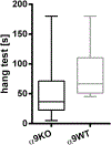
Fig. 1.
Fig. 1.
Mouse hang test. Measurement of hanging time of α9KO and α9WT in seconds (s). α9KO: knockout mice of nicotinic acetylcholine receptor subunit α9; α9WT: corresponding wild type mice of α9KO.
Fig. 2.
Fig. 2.
Bone strength. Measurement of (A) body weight, (B) bending stiffness, (C) maximal breaking force of knockout mice of the α9 and α10 subunits of nicotinic acetylcholine receptors (α9KO, α10KO) and their corresponding wild type mice (WT). * p ≤ 0.05; *** p ≤ 0.001.
Fig. 3.
Fig. 3.
Microarchitecture of mid diaphyseal femur cortex. Calculation of (A) cortical bone area (Ct.Ar), (B) total cross-sectional area inside the periosteal envelope (Tt.Ar), (C) cortical area fraction (Ct.Ar/Tt.Ar), (D) total porosity (Po.tot), (E) 3-dimensional cortical thickness (Ct.Th 3D), (F) tissue mineral density (TMD), and 2D picture of μCT scan of (G) knockout mice of nicotinic acetylcholine receptor subunit α9 (α9KO) and (H) the corresponding wild type mice (α9WT). α10KO: knockout mice of nicotinic acetylcholine receptor subunit α10; α10WT: corresponding wild type mice of α10KO. * p ≤ 0.05; ** p ≤ 0.01. Scale bar: 400 μm.
Fig. 4.
Fig. 4.
Microarchitecture of trabecular bone. Determination of (A) bone volume fraction (BV/TV), (B) trabecular thickness (Tb.Th), (C) trabecular separation (Tb.Sp), (D) specific bone surface (BS/BV), (E) trabecular pattern factor (Tb.Pf), (F) structure model index (SMI) of right distal femur and (G) Tb.Pf, (H) SMI of vertebrae L1, (I) 3D-color coded model of trabecular thickness of α9KO and (J) α9WT distal femur metaphysis. α9KO: knockout mice of nicotinic acetylcholine receptor subunit α9; α10KO: knockout mice of nicotinic acetylcholine receptor subunit α10; α9WT: corresponding wild type mice of α9KO; α10WT: corresponding wild type mice of α10KO. * p ≤ 0.05; ** p ≤ 0.01; *** p ≤ 0.001. (For interpretation of the references to color in this figure legend, the reader is referred to the web version of this article.)
Fig. 5.
Fig. 5.
Osteoblasts. Alkaline phosphatase (ALP) enzyme histochemical staining of (A) α9WT and (B) α10KO. Arrow in B marks single ALP positive osteoblasts. Scale bar: 20 μm. (C) Histomorphometrical calculation of relative ALP perimeter (ALP.Pm/B.Pm) using the ALP enzyme histochemical stainings. mRNA expression analysis of (D) ALP and (E) collagen 1α1 (Col1α1) by means of real-time RT-PCR. Measurement of relative osteoid area (O.Ar/B.Ar) by means of histomorphometrical analysis of Kossa-van-Gieson staining. α9KO: knockout mice of nicotinic acetylcholine receptor subunit α9; α10KO: knockout mice of nicotinic acetylcholine receptor subunit α10; α9WT: corresponding wild type mice of α9KO; α10WT: corresponding wild type mice of α10KO. * p ≤ 0.05.
Fig. 6.
Fig. 6.
Cell culture experiments of neonatal osteoblasts. (A) ALP activity per total protein of α9KO and α9WT cultured in osteogenic differentiation medium (OM) and standard medium (SM). (B) Histomorphometrical analysis of Picro- Sirius-Red positive stained area of α9KO and α9WT osteoblasts in osteogenic differentiation medium. α9KO: knockout mice of nicotinic acetylcholine receptor subunit α9; α9WT: corresponding wild type mice of α9KO. Col 1: collagen type 1, Col 3: collagen type 3. *** p ≤ 0.001.
Fig. 7.
Fig. 7.
Osteocytes. Determination of mRNA expression of (A) connexin 43 (Cx43), (B) SOST, and (C) histomorphometrical quantification of slcerostin im- munopositive osteocytes per bone area (Scl.Ot/B.Ar). Immunohistochemical labeling of osteocytes and their dendrites with (D) antiserum against Cx43 (exemplary picture of α9KO) and (E) sclerostin (exemplarily for α10WT). α9KO: knockout mice of nicotinic acetylcholine receptor subunit α9; α10KO: knockout mice of nicotinic acetylcholine receptor subunit α10; α9WT: corresponding wild type mice of α9KO; α10WT: corresponding wild type mice of α10KO. * p ≤ 0.05. Scale bar: 20 μm.
Fig. 8.
Fig. 8.
Transmission electron microscopy (TEM) of osteocytes. (A) Osteocyte of α9KO with enlarged pericellular space (star), folding of the cytoplasm membrane (arrow), and enlarged space between the two membranes of nucleus (arrow head). (B) Healthy osteocyte of α9WT. (C) Scoring results with significantly increased number of degenerated and dying osteocytes in α9KO compared to α9WT while no significant difference was measured for α10KO and WT. Score 1: healthy, score 2: degenerating, score 3: dying osteocytes. Scale bar: 2 μm.
Fig. 9.
Fig. 9.
Osteoclasts. Histomorphomectical calculation of TRAP stainings of (A) number of osteoclasts per bone perimeter (N.Oc/B.Pm), (B) relative osteoclast perimeter (Oc.Pm/B.Pm) and (C) mRNA expression of Cathepsin K (CtsK) by means of real-time RT-PCR. α9KO: knockout mice of nicotinic acetylcholine receptor subunit α9; α10KO: knockout mice of nicotinic acetylcholine receptor subunit α10; α9WT: corresponding wild type mice of α9KO; α10WT: corresponding wild type mice of α10KO.

References

    1. Mandl P, Hayer S, Karonitsch T, Scholze P, Gyori D, Sykoutri D, Bluml S, Mocsai A, Poor G, Huck S, Smolen JS, Redlich K, Nicotinic acetylcholine receptors modulate osteoclastogenesis, Arthritis Res. Ther 18 (2016) 63. - PMC - PubMed
    1. Bajayo A, Bar A, Denes A, Bachar M, Kram V, Attar-Namdar M, Zallone A, Kovacs KJ, Yirmiya R, Bab I, Skeletal parasympathetic innervation communicates central IL-1 signals regulating bone mass accrual, Proc. Natl. Acad. Sci. U. S. A 109 (2012) 15455–15460. - PMC - PubMed
    1. Lips KS, Kneffel M, Willscheid F, Mathies FM, Kampschulte M, Hartmann S, Panzer I, Durselen L, Heiss C, Kauschke V, Altered ultrastructure, density and cathepsin K expression in bone of female muscarinic acetylcholine receptor M3 knockout mice, Int. Immunopharmacol 29 (2015) 201–207. - PubMed
    1. Kalamida D, Poulas K, Avramopoulou V, Fostieri E, Lagoumintzis G, Lazaridis K, Sideri A, Zouridakis M, Tzartos SJ, Muscle and neuronal nicotinic acetylcholine receptors. Structure, function and pathogenicity, FEBS J. 274 (2007) 3799–3845. - PubMed
    1. Elgoyhen AB, Vetter DE, Katz E, Rothlin CV, Heinemann SF, Boulter J, α10: a determinant of nicotinic cholinergic receptor function in mammalian vestibular and cochlear mechanosensory hair cells, Proc. Natl. Acad. Sci. U. S. A 98 (2001) 3501–3506. - PMC - PubMed

Publication types