

Bi-allelic POLR3A Loss-of-Function Variants Cause Autosomal-Recessive Wiedemann-Rautenstrauch Syndrome
- PMID: 30414627
- PMCID: PMC6288318
- DOI: 10.1016/j.ajhg.2018.10.010
Bi-allelic POLR3A Loss-of-Function Variants Cause Autosomal-Recessive Wiedemann-Rautenstrauch Syndrome
Abstract
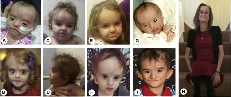
Wiedemann-Rautenstrauch syndrome (WRS), also known as neonatal progeroid syndrome, is a rare disorder of unknown etiology. It has been proposed to be autosomal-recessive and is characterized by variable clinical features, such as intrauterine growth restriction and poor postnatal weight gain, characteristic facial features (triangular appearance to the face, convex nasal profile or pinched nose, and small mouth), widened fontanelles, pseudohydrocephalus, prominent scalp veins, lipodystrophy, and teeth abnormalities. A previous report described a single WRS patient with bi-allelic truncating and splicing variants in POLR3A. Here we present seven additional infants, children, and adults with WRS and bi-allelic truncating and/or splicing variants in POLR3A. POLR3A, the largest subunit of RNA polymerase III, is a DNA-directed RNA polymerase that transcribes many small noncoding RNAs that regulate transcription, RNA processing, and translation. Bi-allelic missense variants in POLR3A have been associated with phenotypes distinct from WRS: hypogonadotropic hypogonadism and hypomyelinating leukodystrophy with or without oligodontia. Our findings confirm the association of bi-allelic POLR3A variants with WRS, expand the clinical phenotype of WRS, and suggest specific POLR3A genotypes associated with WRS and hypomyelinating leukodystrophy.
Keywords: POLR3A, RNA polymerase 3A; Wiedemann-Rautenstrauch syndrome; neonatal progeroid syndrome.
Copyright © 2018 American Society of Human Genetics. Published by Elsevier Inc. All rights reserved.
Figures


References
-
- Rautenstrauch T., Snigula F. Progeria: a cell culture study and clinical report of familial incidence. Eur. J. Pediatr. 1977;124:101–111. - PubMed
-
- Wiedemann H.R. An unidentified neonatal progeroid syndrome: follow-up report. Eur. J. Pediatr. 1979;130:65–70. - PubMed
-
- Pivnick E.K., Angle B., Kaufman R.A., Hall B.D., Pitukcheewanont P., Hersh J.H., Fowlkes J.L., Sanders L.P., O’Brien J.M., Carroll G.S. Neonatal progeroid (Wiedemann-Rautenstrauch) syndrome: report of five new cases and review. Am. J. Med. Genet. 2000;90:131–140. - PubMed
-
- Paolacci S., Bertola D., Franco J., Mohammed S., Tartaglia M., Wollnik B., Hennekam R.C. Wiedemann-Rautenstrauch syndrome: A phenotype analysis. Am. J. Med. Genet. A. 2017;173:1763–1772. - PubMed
-
- Devos E.A., Leroy J.G., Frijns J.P., Van den Berghe H. The Wiedemann-Rautenstrauch or neonatal progeroid syndrome. Report of a patient with consanguineous parents. Eur. J. Pediatr. 1981;136:245–248. - PubMed
Publication types
MeSH terms
Substances
Supplementary concepts
Grants and funding
LinkOut - more resources
Full Text Sources
Other Literature Sources
Medical
Molecular Biology Databases
