. 2019 Jan 1;445(1):90-102.
doi: 10.1016/j.ydbio.2018.11.001. Epub 2018 Nov 8.

Requirement of Pitx2 for skeletal muscle homeostasis

Affiliations

Requirement of Pitx2 for skeletal muscle homeostasis

Chih-Ning Chang et al. Dev Biol. .

Abstract

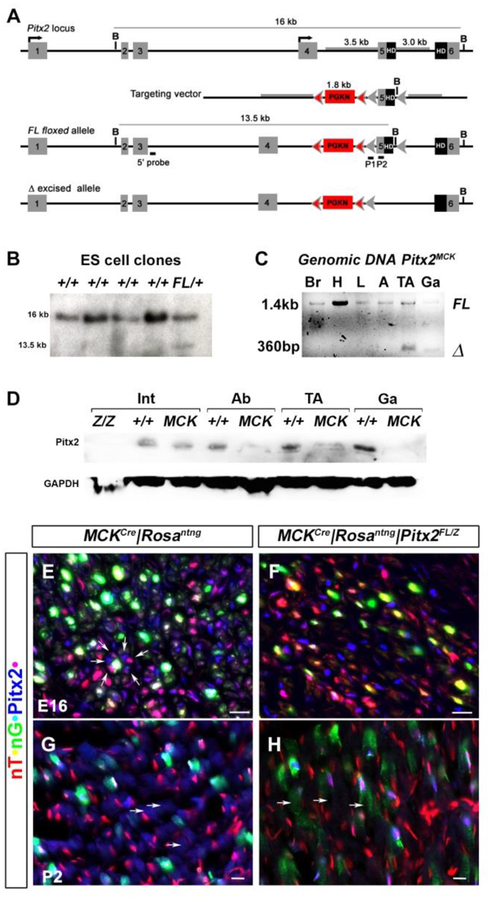
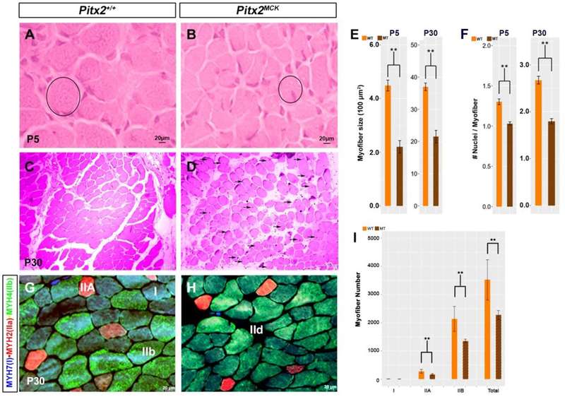
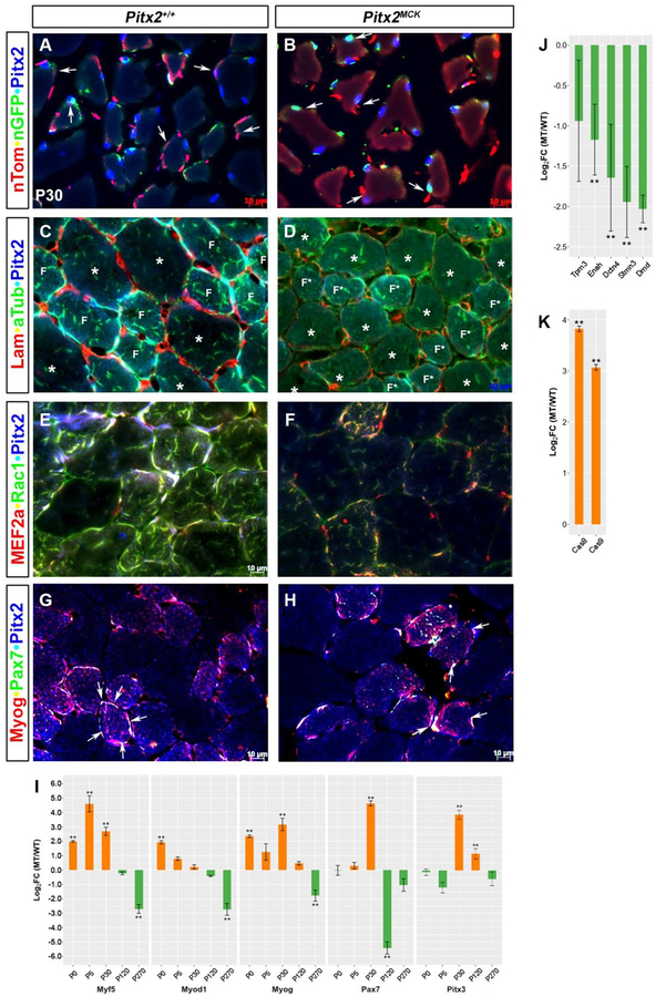
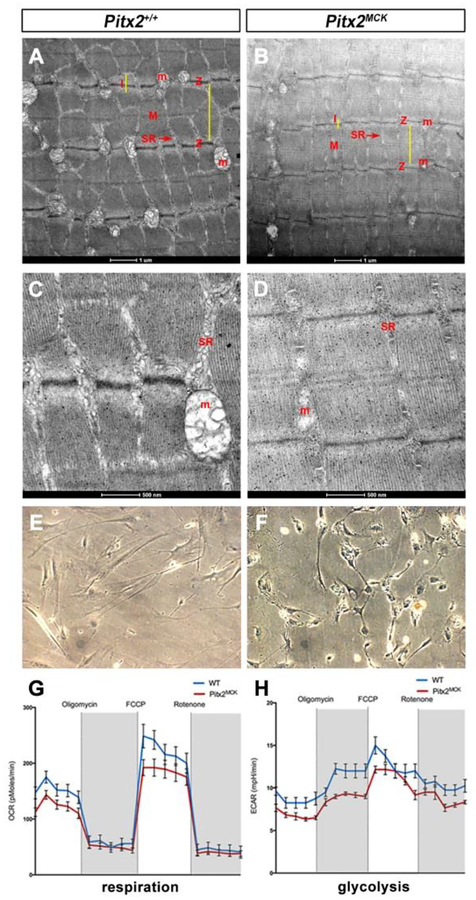
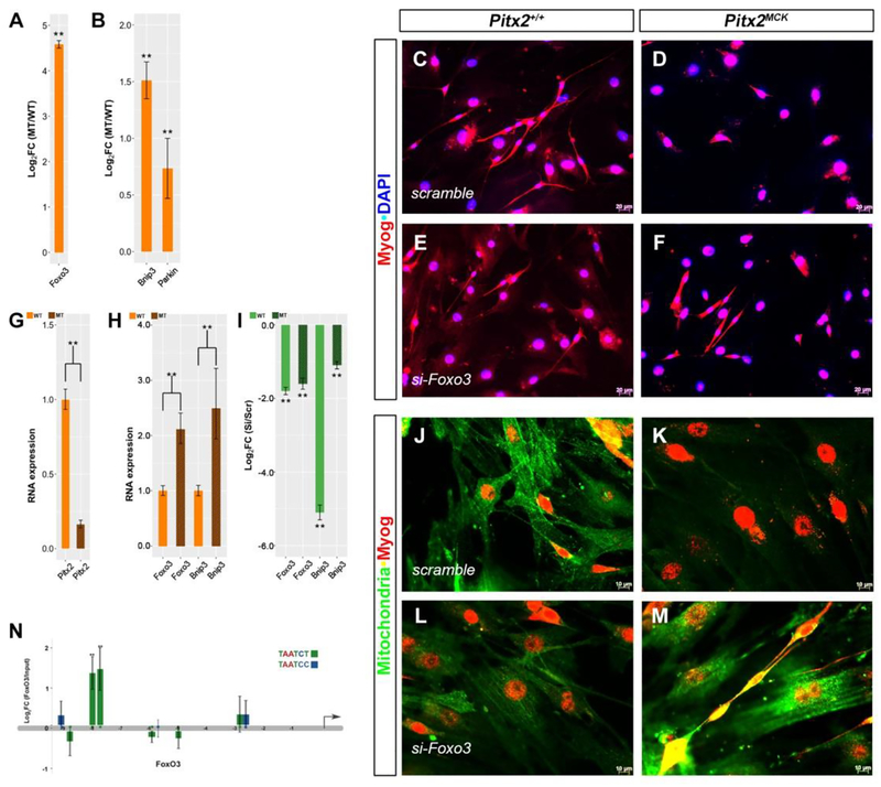
Skeletal muscle is generated by the successive incorporation of primary (embryonic), secondary (fetal), and tertiary (adult) fibers into muscle. Conditional excision of Pitx2 function by an MCKCre driver resulted in animals with histological and ultrastructural defects in P30 muscles and fibers, respectively. Mutant muscle showed severe reduction in mitochondria and FoxO3-mediated mitophagy. Both oxidative and glycolytic energy metabolism were reduced. Conditional excision was limited to fetal muscle fibers after the G1-G0 transition and resulted in altered MHC, Rac1, MEF2a, and alpha-tubulin expression within these fibers. The onset of excision, monitored by a nuclear reporter gene, was observed as early as E16. Muscle at this stage was already severely malformed, but appeared to recover by P30 by the expansion of adjoining larger fibers. Our studies demonstrate that the homeodomain transcription factor Pitx2 has a postmitotic role in maintaining skeletal muscle integrity and energy homeostasis in fetal muscle fibers.

Keywords: Autophagy; Mitochondria; Mouse; Pitx2; Skeletal muscle.


Conflict of interest statement

Competing interests

No competing interests declared.

Figures

Figure 1.
Figure 1.. Generation of new conditional allele and functional confirmation of the Pitx2 muscle specific knockout mouse.
(A) Schematic representations of the endogenous Pitx2 locus, targeting vector, Pitx2FL allele, and the Cre-recombined allele. Exon 5, encoding the upstream end of the homeobox, is flanked by loxP sites (grey arrowheads), while the PGKNeo (PGKN) cassette is flanked by FRT sites (pink arrowheads). Tissue-specific recombination with a Cre driver results in the Pitx2Δ allele (bottom). (B) Homologous recombination in ES cells. Genomic DNA from ES cell clones was digested with BamH1 and probed with the 5’ probe shown above to identify homologous recombination with a13.5 kb fragment (C) Demonstration of Cre recombination of genomic DNA from tissues. The floxed (Pitx2FL) and deleted (Pitx2Δ) form of the allele are detected as 1400 and 360bp amplicons, respectively, in PCR amplifications of genomic DNA isolated from brain (Br), cardiac outflow track (H), liver (L), white adipose tissue (A), or muscles (TA, Ga) of P30 conditional null mice (Pitx2MCK). Excision was only detected tibialis anterior (TA) and gastrocnemius (Ga). (D) Western blot of total protein extracts from the organs of normal and conditional null mice at P30. Pitx2 protein levels were reduced in the abdominal body wall (Ab) and muscles (TA, Ga) of conditional mutants but were unaffected in intestine (I). Intestinal Pitx2 protein levels are reduced in complete (Pitx2Z/Z) but not conditional (Pitx2MCK) mutants. (E-H) Triple labelling immunohistochemistry of E16 hindlimb muscle (E, F) and P2 tibialis anterior (G, H) of control MCKcre|RosanT-nG (E, G) and mutant MCKcre|RosanT-nG |Pitx2FL/Z (F, H) mice. Onset of Cre excision is indicated by the onset of green signal in red nuclei (yellow). Loss of Pitx2 protein signal (blue) was observed in these cells.
Figure 2.
Figure 2.. Atrophying muscle in Pitx2MCK mice.
(A-D) Hematoxylin-eosin staining for TA muscle at P5 (A, B) and P30 (C, D). Myofibers became smaller in diameter (circle), with increased inter-myofiber space, and showed centralized nuclei (arrows) (D). (E) Quantitative comparison of myofiber cross sectional area at P5 and P30. Myofibers of H&E stained cross sections were analyzed using ImageJ. Myofibers of ten sequential sections of the entire TA for both genotypes were counted (n=3). (F) Quantitative analysis of nuclei per myofiber cross section at P5 and P30. (G, H) Triple labelling immunohistochemistry of cross sectioned TA at P30 for MYH7, MYH2 and MYH4 to determine fiber types (I) Quantitative analysis of number of myofibers per area of TA at P30. Three sequential sections of the entire TA for both genotypes were used for the quantitation (n=3).
Figure 3.
Figure 3.. Cytoskeletal Defects Localized to Smaller Fibers in Pitx2MCK muscles.
Immunohistochemical and qPCR comparison of P30 TA muscle Triple labelling to monitor (A, B) docking, conditional allele excision, and Pitx2 expression in myofibers; (C, D) Laminin, alpha-tubulin, and Pitx2 proteins; (E, F) MEF2a, Rac1 and Pitx2 proteins; (G, H) Myog, Pax7, and Pitx2 proteins. Note that smaller (F, F*) and larger(*) fibers respond differently to mutation (see C, D). (I) qPCR analysis of total RNA from TA muscles at P0, P5, P30, P120, and P270. using primers for RNAs encoding muscle specific SSTFs. (J, K) qPCR analysis of P30 TA muscle RNA for cytoskeletal components Tpm3 (Tropomyosin), Enah (Enabled homolog), Dctn4 (Dynactin), Stmn3 (Stathmin), Dmd (Dystrophin), and apoptosis proteins (Caspases 8 and 9).
Figure 4.
Figure 4.. Mitochondrial Loss and Respiration Defects in Pitx2MCK Muscle.
(A, D) Transmission electron microscopy for TA at P30 indicated distorted and smaller sarcomeres. Z, I, and M bands show differences. Mitochondria (M) and sarcoplasmic reticulum (SR) are vestigial. (E-H) Seahorse assay on primary TA muscle cell cultures (E, F) from WT and Pitx2MCK mice (n=3). (G) OCR and (H) ECAR measure mitochondrial respiration and glycolysis, respectively.
Figure 5.
Figure 5.. Pitx2 Suppresses FoxO3-dependent Mitophagy.
(A, B) RT-qPCR of total RNA from P30 TA muscle of WT and Pitx2MCK mice. (C-F) Cultured myocytes from P30 TA myofibers from normal (C, E, J, L) and mutant (D, F, K, M) were transfected with scramble siRNA (C, D, J, K) and Foxo3 siRNA (E, F, L, M). Cells were stained for Myog and DAPI (C-F) or Myog and Mitochondria (J-M). (G, H) qPCR of total RNA from scramble treated cell cultures, indicated reduced Pitx2 and increased Foxo3 and Bnip3 levels in mutant cultures (I) qPCR analysis comparing expression between scramble and si-FoxO3 treatments. Levels of FoxO3 and Bnip3 RNAs decline with si-FoxO3 treatment. (N) RT-qPCR ChIP for Pitx2 occupancy on 10 Kb upstream of the FoxO3 transcription start site. Pitx2 bicoid binding domains TAATCT (green) and TAATCC (blue) indicated Pitx2 occupancy (TAATCY; −9170, −8876, −8102, −7816, −6013, −5877, −5099, −2950, −2739). Pitx2 occupancy was greatest at positions −8102, −7816.

References

    1. Abu-Elmagd M, Robson L, Sweetman D, Hadley J, Francis-West P, Münsterberg A, 2010. Wnt/Lef1 signaling acts via Pitx2 to regulate somite myogenesis. Dev. Biol 337, 211–219. doi:10.1016/j.ydbio.2009.10.023 - DOI - PubMed
    1. Ai D, Liu W, Ma L, Dong F, Lu M-F, Wang D, Verzi MP, Cai C, Gage PJ, Evans S, Black BL, Brown NA, Martin JF, 2006. Pitx2 regulates cardiac left-right asymmetry by patterning second cardiac lineage-derived myocardium. Developmental Biology 296, 437–449. doi:10.1016/j.ydbio.2006.06.009 - DOI - PMC - PubMed
    1. Alvares LE, Schubert FR, Thorpe C, Mootoosamy RC, Cheng L, Parkyn G, Lumsden A, Dietrich S, 2003. Intrinsic, Hox-dependent cues determine the fate of skeletal muscle precursors. DEVCEL 5, 379–390. - PubMed
    1. Bajanca F, Luz M, Raymond K, Martins GG, Sonnenberg A, Tajbakhsh S, Buckingham M, Thorsteinsdóttir S, 2006. Integrin alpha6beta1-laminin interactions regulate early myotome formation in the mouse embryo. Development 133, 1635–1644. doi:10.1242/dev.02336 - DOI - PubMed
    1. Berkes CA, Tapscott SJ, 2005. MyoD and the transcriptional control of myogenesis. Seminars in Cell and Developmental Biology 16, 585–595. doi:10.1016/j.semcdb.2005.07.006 - DOI - PubMed

Publication types