

Molecular biomarkers of Graves' ophthalmopathy
- PMID: 30414981
- PMCID: PMC6381289
- DOI: 10.1016/j.yexmp.2018.11.004
Molecular biomarkers of Graves' ophthalmopathy
Abstract
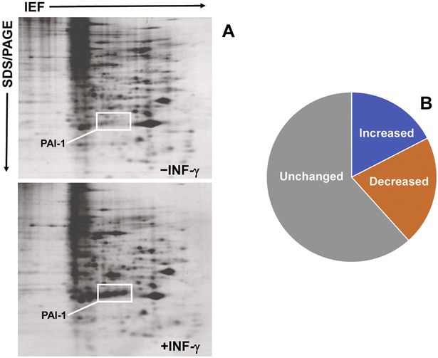
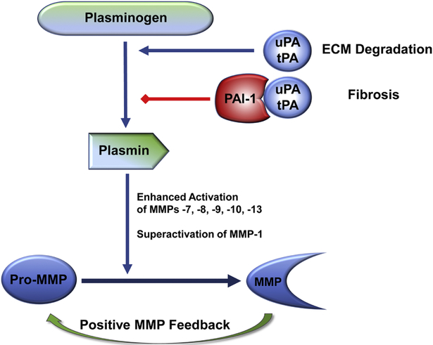
Graves' ophthalmopathy (GO), a complication of Graves' disease (GD), is typified by orbital inflammation, ocular tissue expansion and remodeling and, ultimately, fibrosis. Orbital fibroblasts are key effectors of GO pathogenesis exhibiting exaggerated inflammatory and fibroproliferative responses to cytokines released by infiltrating immune cells. Activated orbital fibroblasts also produce inflammatory mediators that contribute to disease progression, facilitate the orbital trafficking of monocytes and macrophages, promote differentiation of matrix-producing myofibroblasts and stimulate accumulation of a hyaluronan-rich stroma, which leads to orbital tissue edema and fibrosis. Proteomic and transcriptome profiling of the genomic response of ocular and non-ocular fibroblasts to INF-γ and TGF-β1 focused on identification of translationally-relevant therapeutic candidates. Induction of plasminogen activator inhibitor-1 (PAI-1, SERPINE1), a clade E member of the serine protease inhibitor (SERPIN) gene family and a prominent regulator of the pericellular proteolytic microenvironment, was one of the most highly up-regulated proteins in INF-γ- or TGF-β1-stimulated GO fibroblasts as well as in severe active GD compared to patients without thyroid disease. PAI-1 has multifunctional roles in inflammatory and fibrotic processes that impact tissue remodeling, immune cell trafficking and survival as well as signaling through several receptor systems. This review focuses on the pathophysiology of the GO fibroblast and possible targets for effective drug therapy.
Keywords: Biomarkers; Fibrosis; Graves' disease; Inflammatory cytokines; Orbitopathy; PAI-1; Plasmin cascade; SERPINs; TGF-β; Tissue remodeling.
Copyright © 2018 Elsevier Inc. All rights reserved.
Conflict of interest statement
Conflict of interest
The authors declare no conflict of interest.
Figures
References
-
- Anderson RL, Tweeten JP, Patrinely JR, Garland PE, Thiese SM, 1989. Dysthyroid optic neuropathy without extraocular muscle involvement. Ophthalmic Surg. 20, 568–574. - PubMed
-
- Aniszewski JP, Valyasevi RW, Bahn RS, 2000. Relationship between disease duration and predominant orbital T cell subset in Graves' ophthalmopathy. J. Clin. Endocrinol. Metab 85, 776–780. - PubMed
-
- Aso Y, 2007. Plasminogen activator inhibitor (PAI)-1 in vascular inflammation and thrombosis. Front. Biosci 12, 2957–2966. - PubMed
-
- Bahn RS, 2003. Clinical review 157: pathophysiology of Graves' ophthalmopathy: the cycle of disease. J. Clin. Endocrinol. Metab 88, 1939–1946. - PubMed
Publication types
MeSH terms
Substances
Grants and funding
LinkOut - more resources
Full Text Sources
Miscellaneous
