. 2018 Nov 7;26(11):2542-2552.
doi: 10.1016/j.ymthe.2018.09.008. Epub 2018 Sep 13.

Low-Dose Radiation Conditioning Enables CAR T Cells to Mitigate Antigen Escape

Affiliations

Low-Dose Radiation Conditioning Enables CAR T Cells to Mitigate Antigen Escape

Carl DeSelm et al. Mol Ther. .

Abstract

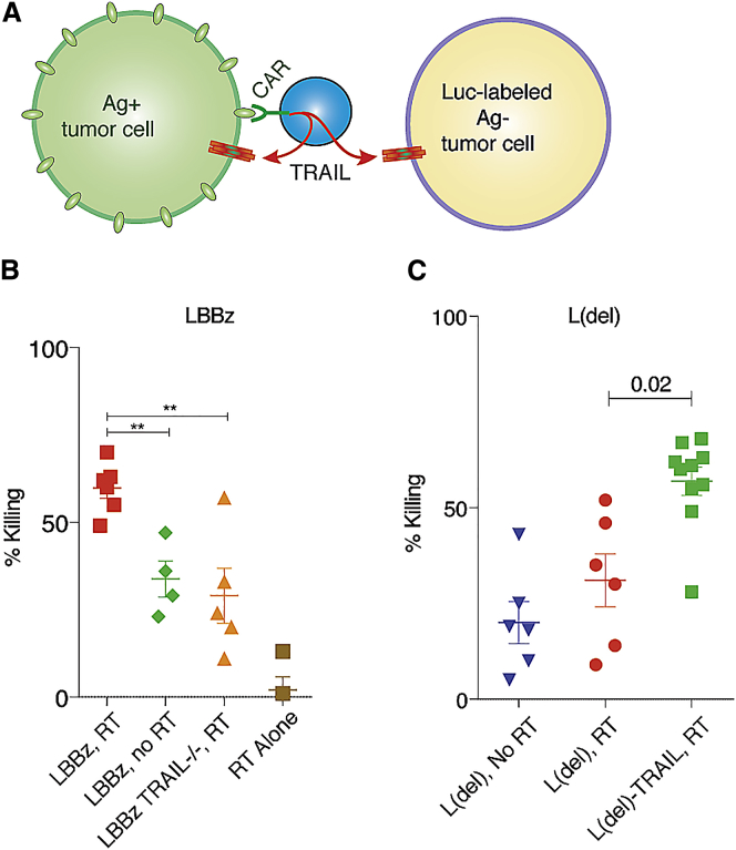
CD19 chimeric antigen receptors (CARs) have demonstrated great efficacy against a range of B cell malignancies. However, antigen escape and, more generally, heterogeneous antigen expression pose a challenge to applying CAR therapy to a wide range of cancers. We find that low-dose radiation sensitizes tumor cells to immune rejection by locally activated CAR T cells. In a model of pancreatic adenocarcinoma heterogeneously expressing sialyl Lewis-A (sLeA), we show that not only sLeA+ but also sLeA- tumor cells exposed to low-dose radiation become susceptible to CAR therapy, reducing antigen-negative tumor relapse. RNA sequencing analysis of low-dose radiation-exposed tumors reveals the transcriptional signature of cells highly sensitive to TRAIL-mediated death. We find that sLeA-targeted CAR T cells produce TRAIL upon engaging sLeA+ tumor cells, and eliminate sLeA- tumor cells previously exposed to systemic or local low-dose radiation in a TRAIL-dependent manner. These findings enhance the prospects for successfully applying CAR therapy to heterogeneous solid tumors. Local radiation is integral to many tumors' standard of care and can be easily implemented as a CAR conditioning regimen.

Keywords: CAR T cell; antigen escape; pancreatic cancer; radiation; sialyl Lewis-A.


Figures

None
Graphical abstract
Figure 1
Figure 1
RT Sensitizes Pancreatic Cancer to CAR T Cell Killing without Affecting Target Antigen Expression (A) Tumor cell viability 48 hr after exposure to various doses of radiation. (B) Capan2 pancreatic cancer cells were exposed to low-dose RT (2 Gy) and, 48 hr later, incubated with CAR T cells at the indicated ratios for 18 hr, after which percent killing was determined. (C) Target antigen expression levels were unchanged 48 hr after RT. (D) Transcriptome analysis of target cells 6 hr after RT reveals a number of significantly affected apoptotic pathways. (E) TRAIL mRNA expression and protein levels in the media of CAR T cells after exposure to target antigen (sLeA-expressing Capan2 cells). (F) TRAIL protein was quantified in the media of LBBz and L(del) CAR T cells grown on target cells expressing or not expressing the target antigen. LFC, log2 fold change; E:T, effector:target. Error bars = SEM.
Figure 2
Figure 2
TRAIL Expressed by Activated CAR T Cells Is Active against Antigen-Negative Tumor Cells in a Heterogeneous Tumor Population Exposed to Low-Dose Radiation (A) CAR-activated T cells produce TRAIL, which acts upon radiation-sensitized antigen-positive and antigen-negative tumor cells. (B and C) Ag+ cells were mixed with luciferase-expressing Ag cells at a ratio of 75:25, exposed to low-dose RT, and cocultured with LBBz (B) or L(del) (C) CAR T cells for 4 days, followed by luciferase-based quantification of cell killing. **p < 0.01.
Figure 3
Figure 3
Sensitizing RT Transcriptionally Primes Pancreatic Cancer Cells for TRAIL-Induced Death (A) RNA expression levels of signaling molecules known to mediate various TRAIL responses, including survival and migration, tumor-supportive inflammation, necroptosis, apoptosis, and death receptor endocytosis, were quantified by RNA-seq before and after RT exposure to Capan2 pancreatic cancer cells in three biologic replicates. Significantly induced and downregulated molecules are shown in red and green, respectively, with magnitude represented by color gradient. Molecules in gray were not significantly changed. (B) CTV-labeled Ag cells were exposed to RT 2 days before coculture with unlabeled Ag+ cells, annexin-V 595, and TRAIL−/− or TRAILWT CAR T cells. Cultures were monitored by live video microscopy, and Ag cell apoptosis was quantified over time. Error bars = SEM.
Figure 4
Figure 4
Sensitizing RT Allows CAR T Cells to Eliminate Heterogeneous PDAC In Vivo (A) Capan2 tumor cells were mixed at 75:25 sLeA+:sLeA and then injected into the pancreas of NSG mice. After tumors established for 9 days, mice were given RT, followed by CAR T cells. (B) Waterfall plot of tumor volume change at time of death among different treatment groups. (C–H) BLI was performed weekly on mice that were untreated (C) or treated with RT and 1928z (D), LBBz (E), RT and LBBz (F), RT and TCR−/− LBBz (G), or RT and TRAIL−/− LBBz (H) CAR T cells. (I–K) T cell infiltration of tumors from CAR-treated or RT with CAR-treated mice was determined using BLI T cell imaging (detecting G-Luc on the transduced T cell) over the first 19 days (I) and by IHC from mice sacrificed on day 21 (J and K, all not significant [ns]). (L) Tumors in mice that progressed displayed reduced target antigen expression over time by FACS. (M) BLI of mice treated with RT+L(del) or RT+L(del)-TRAIL CAR T cells. Error bars = SEM.
Figure 5
Figure 5
Outcome of a Large-Cell Lymphoma Patient with a Heterogeneous Tumor Treated with Palliative RT and CD19 CAR T Cells (A) Total body or local RT was delivered to mice harboring heterogeneous tumors of the pancreas using image-guided radiation, followed by CAR T cells. (B) Tumor burden was monitored by BLI. (C and D) Patient biopsy before CAR T cell treatment examined for CD19 by IHC (C) and flow cytometry (D). (E) Fluorodeoxyglucose (FDG)-PET scan before and 1, 2, and 6 months after palliative leg RT and systemic 1928z CAR T cells. Error bars = SEM.

References

    1. Sadelain M., Rivière I., Riddell S. Therapeutic T cell engineering. Nature. 2017;545:423–431. - PMC - PubMed
    1. June C.H., Sadelain M. Chimeric Antigen Receptor Therapy. N Engl J Med. 2018;379:64–73. - PMC - PubMed
    1. Brentjens R.J., Latouche J.B., Santos E., Marti F., Gong M.C., Lyddane C., King P.D., Larson S., Weiss M., Rivière I., Sadelain M. Eradication of systemic B-cell tumors by genetically targeted human T lymphocytes co-stimulated by CD80 and interleukin-15. Nat. Med. 2003;9:279–286. - PubMed
    1. Fry T.J., Shah N.N., Orentas R.J., Stetler-Stevenson M., Yuan C.M., Ramakrishna S., Wolters P., Martin S., Delbrook C., Yates B. CD22-targeted CAR T cells induce remission in B-ALL that is naive or resistant to CD19-targeted CAR immunotherapy. Nat. Med. 2018;24:20–28. - PMC - PubMed
    1. Wilkie S., van Schalkwyk M.C., Hobbs S., Davies D.M., van der Stegen S.J., Pereira A.C., Burbridge S.E., Box C., Eccles S.A., Maher J. Dual targeting of ErbB2 and MUC1 in breast cancer using chimeric antigen receptors engineered to provide complementary signaling. J. Clin. Immunol. 2012;32:1059–1070. - PubMed

Publication types

MeSH terms