. 2019 Jan;152(1):11-19.
doi: 10.1016/j.ygyno.2018.10.032. Epub 2018 Nov 8.

Analysis of mutational signatures in primary and metastatic endometrial cancer reveals distinct patterns of DNA repair defects and shifts during tumor progression

Affiliations

Analysis of mutational signatures in primary and metastatic endometrial cancer reveals distinct patterns of DNA repair defects and shifts during tumor progression

Charles W Ashley et al. Gynecol Oncol. 2019 Jan.

Abstract

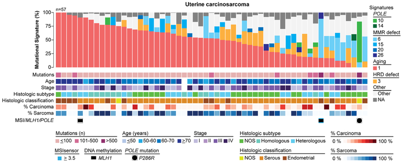
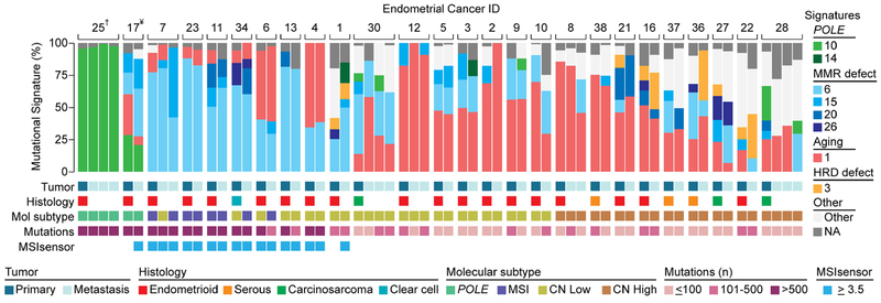
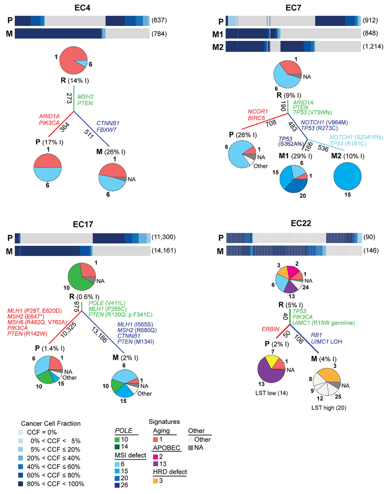
Objective: Mutational signatures provide insights into the biological processes shaping tumor genomes and may inform patient therapy. We sought to define the mutational signatures of i) endometrioid and serous endometrial carcinomas (ECs), stratified into the four molecular subtypes, ii) uterine carcinosarcomas, and iii) matched primary and metastatic ECs.

Methods: Whole-exome sequencing MC3 data from primary endometrioid and serous carcinomas (n = 232) and uterine carcinosarcomas (n = 57) from The Cancer Genome Atlas (TCGA), and matched primary and metastatic ECs (n = 61, 26 patients) were reanalyzed, subjected to mutational signature analysis using deconstructSigs, and correlated with clinicopathologic and genomic data.

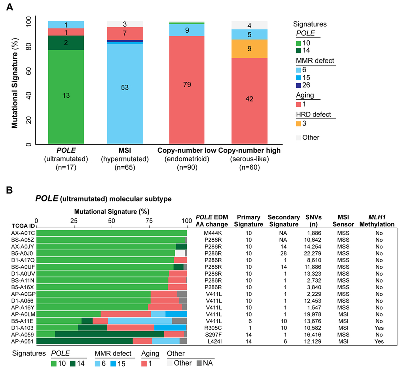
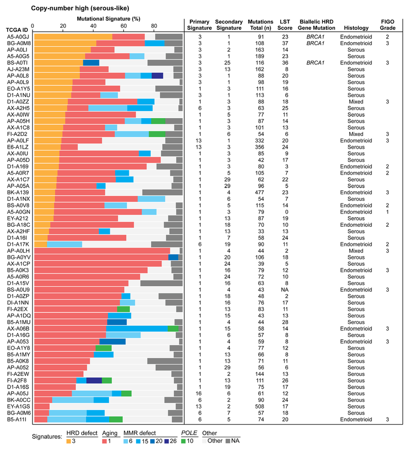
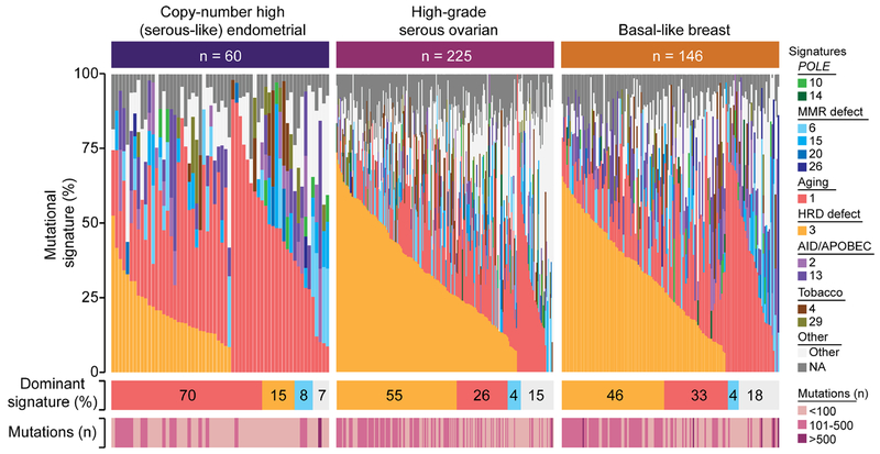
Results: POLE (ultramutated) and MSI (hypermutated) molecular subtypes displayed dominant mutational signatures associated with POLE mutations (15/17 cases) and microsatellite instability (55/65 cases), respectively. Most endometrioid and serous carcinomas of copy-number low (endometrioid) and copy-number high (serous-like) molecular subtypes, and carcinosarcomas displayed a dominant aging-associated signature 1. Only 15% (9/60) of copy-number high (serous-like) ECs had a dominant signature 3 (homologous recombination DNA repair deficiency (HRD)-related), a prevalence significantly lower than that found in high-grade serous ovarian carcinomas (54%, p < 0.001) or basal-like breast cancers (46%, p < 0.001). Shifts from aging- or POLE- to MSI-related mutational processes were observed in the progression from primary to metastatic ECs in a subset of cases.

Conclusions: The mutational processes underpinning ECs vary even among tumors of the same TCGA molecular subtype and in the progression from primary to metastatic ECs. Only a minority of copy-number high (serous-like) ECs display genomics features of HRD and would likely benefit from HRD-directed therapies.

Keywords: Carcinosarcoma; Endometrial cancer; Metastasis; Molecular subtypes; Mutational signatures.


Conflict of interest statement

CONFLICT OF INTEREST

N. Riaz reports research support from Pfizer and Bristol-Myers Squibb, and personal fees from the Illumina Speakers’ Bureau, outside the submitted work. J.S. Reis-Filho reports personal/consultancy fees from VolitionRx, Page.AI and Goldman Sachs, outside the submitted work. The remaining authors have no conflicts of interest to declare.

Figures

Figure 1.
Figure 1.. Mutational signatures of primary endometrioid and serous endometrial cancers stratified according to molecular subtype.
A) The dominant mutational signatures of 232 endometrioid and serous endometrial carcinomas from The Cancer Genome Atlas (TCGA) [3] stratified according to molecular subtype. Mutational signatures are color-coded according to the legend on the right, and the number of endometrial cancers displaying a specific dominant mutational signature is shown in each bar. B) The mutational signatures of the 17 endometrial cancers of the 232 endometrioid and serous endometrial cancers studied by TCGA [3] classified as of POLE (ultramutated) molecular subtype, harboring POLE exonuclease domain mutations, are shown. Cases are sorted by their proportion of signature 10 associated with POLE alterations, and molecular signatures are color-coded according to the legend. Information on the exact POLE exonuclease domain mutation (amino acid), the primary and secondary molecular signatures, the number of non-synonymous single nucleotide variants (SNVs), microsatellite instability (MSIsensor score) and MLH1 promoter methylation is provided for each case on the right. AA, amino acid change; EDM, exonuclease domain mutation; MSI, microsatellite instability; MSS, microsatellite stable.
Figure 2.
Figure 2.. Mutational signatures and homologous recombination DNA repair defects in endometrial cancers of copy-number high (serous-like) molecular subtype.
Mutational signatures of 60 endometrioid and serous endometrial carcinomas of copy-number high (serous-like) molecular subtype from The Cancer Genome Atlas (TCGA), sorted according to the proportion of mutational signatures 3 and 1. Mutational signatures are color-coded according to the legend on the bottom. Information on the primary and secondary signatures, total number of somatic mutations, large-scale state transition (LST) scores, bi-allelic homologous recombination DNA repair (HRD) gene mutations, histology and FIGO grade for endometrioid and mixed endometrial carcinomas are provided for each case on the right.
Figure 3.
Figure 3.. Mutational signature 3 associated with homologous recombination DNA repair defects in copy-number high (serous-like) endometrial cancers, high-grade serous ovarian cancers and basal-like breast cancers from The Cancer Genome Atlas.
Mutational signatures were defined for copy-number high (serous-like) endometrial cancers (n=60), high-grade serous ovarian cancers (n=225) and basal-like breast cancers (n=146) with ≥20 single nucleotide variants from The Cancer Genome Atlas (TCGA) [3,15,16], color-coded according to the legend. Cases are sorted by the proportion of signatures 3 and 1. The percentage of each dominant signature and the total number of non-synonymous somatic mutations for each case are shown below. HRD, homologous recombination DNA repair deficiency; MSI, microsatellite instability.
Figure 4.
Figure 4.. Mutational signatures of uterine carcinosarcomas.
Mutational signatures for 57 primary treatment-naïve uterine carcinosarcomas from The Cancer Genome Atlas (TCGA) [5], sorted by the proportion of signature 1, and color-coded according to the legend. The number of non-synonymous somatic mutations per case, age at diagnosis, stage, histologic subtype, histologic classification, percentage of carcinoma in the frozen tissue subjected to whole-exome sequencing, percentage of sarcoma in the frozen tissue subjected to whole-exome sequencing, MSIsensor score, MLH1 promoter methylation and POLE mutations are provided below the signatures. HRD, homologous recombination DNA repair deficiency; MSI, microsatellite instability; NOS, not otherwise specified.
Figure 5.
Figure 5.. Mutational signatures of primary endometrial cancers and matched metastases.
Mutational signatures of primary and metastatic endometrial cancers from 26 patients from Gibson et al [6], sorted according to molecular subtype, color-coded according to the legend. Information on histology and molecular subtype was obtained from Gibson et al [6] and is displayed below each case along with the number of non-synonym ous somatic mutations and MSIsensor scores. HRD, homologous recombination DNA repair deficiency; MSI, microsatellite instability, †, P286R POLE somatic mutation; ¥, V411L POLE somatic mutation.
Figure 6.
Figure 6.. Mutational signatures of shared and private mutations identified in primary endometrial cancers and matched metastases.
Cancer cell fractions of non-synonymous somatic mutations identified in primary endometrial cancers and metastases using ABSOLUTE [26], color-coded according to the legend. The number of non-synonymous somatic mutations identified in a given lesion is shown in parentheses on the right. Pie charts represent the mutational signatures identified in mutations shared between primary tumors and metastases (root, R) in mutations private to the primary lesion (P) and in mutations private to the metastases (M), color-coded according to the legend. The length of the branches is proportional to the number of somatic mutations that are shared/ unique to a given lesion, and the number of mutations is shown alongside the branches. The percentage of small insertions and deletions (indels, I) are included within parentheses next the R, P or M labels. Selected pathogenic somatic mutations are shown along their corresponding branches. For EC22, the LST scores are shown below the pie charts. EC, endometrial cancer; LST, large-scale state transition; M, metastasis; P, primary tumor; R, root. Additional plots are shown in Supplementary Fig. S5.

Comment in

References

    1. Siegel RL, Miller KD, Jemal A. Cancer statistics, 2018. CA Cancer J Clin. 68 (2018) 7–30. - PubMed
    1. Jemal A, Ward EM, Johnson CJ, Cronin KA, Ma J, Ryerson B, et al. Annual Report to the Nation on the Status of Cancer, 1975-2014, Featuring Survival. J Natl Cancer Inst. 109 (2017). - PMC - PubMed
    1. Cancer Genome Atlas Research Network, Kandoth C, Schultz N, Cherniack AD, Akbani R, Liu Y, et al. Integrated genomic characterization of endometrial carcinoma. Nature. 497 (2013) 67–73. - PMC - PubMed
    1. Cosgrove CM, Tritchler DL, Cohn DE, Mutch DG, Rush CM, Lankes HA, et al. An NRG Oncology/GOG study of molecular classification for risk prediction in endometrioid endometrial cancer. Gynecol Oncol. 148 (2018) 174–180. - PMC - PubMed
    1. Cherniack AD, Shen H, Walter V, Stewart C, Murray BA, Bowlby R, et al. Integrated Molecular Characterization of Uterine Carcinosarcoma. Cancer Cell. 31 (2017) 411–423. - PMC - PubMed

Publication types