

Signal Intensity of the Cerebrospinal Fluid after Intravenous Administration of Gadolinium-based Contrast Agents: Strong Contrast Enhancement around the Vein of Labbe
- PMID: 30416181
- PMCID: PMC6630048
- DOI: 10.2463/mrms.mp.2018-0043
Signal Intensity of the Cerebrospinal Fluid after Intravenous Administration of Gadolinium-based Contrast Agents: Strong Contrast Enhancement around the Vein of Labbe
Abstract
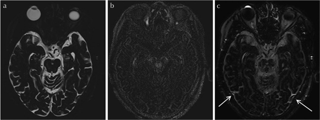
Purpose: Since the first report on the deposition of gadolinium in the brain parenchyma after repeated intravenous administrations of gadolinium-based contrast agent GBCA (IV-GBCA), the mechanisms of penetration and retention are still remaining a hot topic of discussion and a target of investigation. We routinely obtain endolymphatic hydrops (EH) images at 4 h after IV administration of a single dose (SD) of GBCA (IV-SD-GBCA) using heavily T2-weighted three-dimensional fluid-attenuated inversion recovery imaging (hT2W-3D-FLAIR). Occasionally, we have encountered cases, which indicate high-signal intensity (SI) in the cerebrospinal fluid (CSF) surrounding the vein of Labbe. The purpose of the present study was to investigate the degree of contrast enhancement of the CSF surrounding the vein of Labbe on hT2W-3D-FLAIR after IV-SD-GBCA in comparison with other CSF spaces.
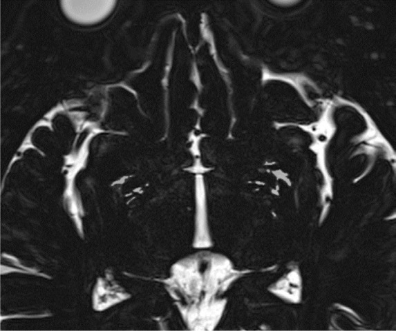
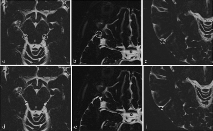
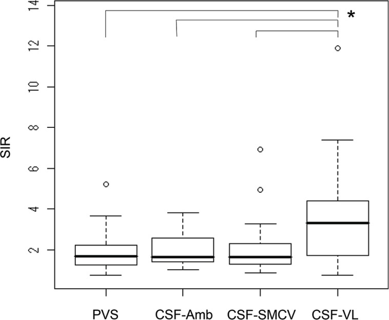
Materials and methods: In 25 patients with a suspicion of EH, a magnetic resonance cisternography (MRC) and an hT2W-3D-FLAIR were obtained at 4 h after IV-SD-GBCA. The perivascular space (PVS) in the basal ganglia, CSF spaces in the ambient cistern (CSF-Amb), the CSF surrounding the superficial middle cerebral vein (CSF-SMCV), and the CSF surrounding the vein of Labbe (CSF-VL) were segmented on MRC. The PVS and CSF regions were co-registered onto the hT2W-3D-FLAIR and the SI of the PVS and CSF spaces were measured. The SI ratio (SIR) of the post-contrast hT2W-3D-FLAIR to the pre-contrast hT2W-3D-FLAIR was measured. Significant differences were evaluated using Steel-Dwass's test for multiple comparisons.
Results: The SIR of the CSF-VL was significantly higher than that of the PVS (P = 0.008), the CSF-Amb (P = 0.021), and the CSF-SMCV (P = 0.023).
Conclusion: The strong contrast enhancement of CSF space around the vein of Labbe was confirmed on hT2W-3D-FLAIR at 4 h after IV-GBCA compared to the PVS and the other CSF spaces.
Keywords: cerebrospinal fluid; gadolinium; glymphatic system; magnetic resonance imaging; vein.
Conflict of interest statement
All authors do not have any conflicts of interest regarding the present study.
Figures
Similar articles
-
Relationship between Contrast Enhancement of the Perivascular Space in the Basal Ganglia and Endolymphatic Volume Ratio.Magn Reson Med Sci. 2018 Jan 10;17(1):67-72. doi: 10.2463/mrms.mp.2017-0001. Epub 2017 Jun 8. Magn Reson Med Sci. 2018. PMID: 28592709 Free PMC article.
-
Gd-based Contrast Enhancement of the Perivascular Spaces in the Basal Ganglia.Magn Reson Med Sci. 2017 Jan 10;16(1):61-65. doi: 10.2463/mrms.mp.2016-0039. Epub 2016 Jul 12. Magn Reson Med Sci. 2017. PMID: 27430361 Free PMC article.
-
Association between the Putative Meningeal Lymphatics at the Posterior Wall of the Sigmoid Sinus and Delayed Contrast-agent Elimination from the Cerebrospinal Fluid.Magn Reson Med Sci. 2024 Jan 1;23(1):80-91. doi: 10.2463/mrms.mp.2022-0110. Epub 2023 Jan 19. Magn Reson Med Sci. 2024. PMID: 36653154 Free PMC article.
-
Technical considerations for using intravenous gadolinium-based-contrast-agent (GBCA) based MRI approaches to study cerebrospinal fluid (CSF) circulation and clearance.Neuroimage. 2025 May 15;312:121239. doi: 10.1016/j.neuroimage.2025.121239. Epub 2025 Apr 24. Neuroimage. 2025. PMID: 40280217 Review.
-
Optimal sequences and sequence parameters for GBCA-enhanced MRI of the glymphatic system: a systematic literature review.Acta Radiol. 2021 Oct;62(10):1324-1332. doi: 10.1177/0284185120969950. Epub 2020 Nov 5. Acta Radiol. 2021. PMID: 33153270
Cited by
-
Neurofluid Dynamics and the Glymphatic System: A Neuroimaging Perspective.Korean J Radiol. 2020 Nov;21(11):1199-1209. doi: 10.3348/kjr.2020.0042. Epub 2020 Aug 11. Korean J Radiol. 2020. PMID: 32783417 Free PMC article. Review.
-
The Regulation of Cerebral Spinal Fluid Flow and Its Relevance to the Glymphatic System.Curr Neurol Neurosci Rep. 2020 Oct 19;20(12):58. doi: 10.1007/s11910-020-01077-9. Curr Neurol Neurosci Rep. 2020. PMID: 33074399 Free PMC article. Review.
-
Automated Methods for Detecting and Quantitation of Enlarged Perivascular spaces on MRI.J Magn Reson Imaging. 2023 Jan;57(1):11-24. doi: 10.1002/jmri.28369. Epub 2022 Jul 22. J Magn Reson Imaging. 2023. PMID: 35866259 Free PMC article. Review.
-
The "hype" of hydrops in classifying vestibular disorders: a narrative review.J Neurol. 2020 Dec;267(Suppl 1):197-211. doi: 10.1007/s00415-020-10278-8. Epub 2020 Nov 17. J Neurol. 2020. PMID: 33201310 Free PMC article. Review.
-
Glymphatic-lymphatic coupling: assessment of the evidence from magnetic resonance imaging of humans.Cell Mol Life Sci. 2024 Mar 13;81(1):131. doi: 10.1007/s00018-024-05141-2. Cell Mol Life Sci. 2024. PMID: 38472405 Free PMC article. Review.
References
-
- Weinmann HJ, Brasch RC, Press WR, Wesbey GE. Characteristics of gadolinium-DTPA complex: a potential NMR contrast agent. AJR Am J Roentgenol 1984; 142:619–624. - PubMed
-
- Naganawa S, Suzuki K, Yamazaki M, Sakurai Y. Serial scans in healthy volunteers following intravenous administration of gadoteridol: time course of contrast enhancement in various cranial fluid spaces. Magn Reson Med Sci 2014; 13:7–13. - PubMed
MeSH terms
Substances
LinkOut - more resources
Full Text Sources
Research Materials