. 2018 Oct 15;10(10):3211-3223.
eCollection 2018.

The FENDRR/miR-214-3P/TET2 axis affects cell malignant activity via RASSF1A methylation in gastric cancer

Affiliations

The FENDRR/miR-214-3P/TET2 axis affects cell malignant activity via RASSF1A methylation in gastric cancer

Zhaocai He et al. Am J Transl Res. .

Abstract

To explore the effect of fetal-lethal non-coding developmental regulatory RNA (FENDRR) in the initiation and progression of gastric cancer (GC). We detected the levels of FENDRR, microRNA-214-3p (miR-214-3p), and ten-eleven-translocation (TET2) in GC tissues and GC cell lines. In addition, we evaluated the location of FENDRR in GC cell lines by fluorescence in situ hybridization (FISH). Cell proliferation and apoptosis were measured by CCK-8 and Hoechst staining assays. Methylation-specific PCR assay (MSP) was used to evaluate the methylation status of ras-association domain family 1A (RASSF1A). We also observed the direct binding of miR-214-3p on FENDRR by dual-luciferase activity assay in GC cells. FENDRR and TET2 expressions were significantly down-regulated and miR-214-3p was up-regulated in gastric cancer tissues compared to adjacent unaffected tissues. In addition, RASSF1A was hypermethylated in gastric cancer tissues compared to adjacent tissues. The expressions of all the three indicators were influenced by differentiation of tumor, TNM stage of tumors, and lymph node metastasis in patients with GC. A gastric cancer cell line with low FENDRR expression compared to a high FENDRR expressing cell line showed again increased miR-214-3p expression, decreased TET2 and RASSF1A expressions, and RASSF1A hypermethylation, resulting in decreased apoptosis and increased proliferation. Furthermore, we observed a negative correlation between FENDRR and miR-214-3p in GC. The FENDRR/miR-214-3P/TET2 axis plays a critical role in GC progress via methylation of RASSF1A.

Keywords: FENDRR; RASSF1A; TET2; gastric cancer; methylation; miR-214-3p; proliferation.


Conflict of interest statement

None.

Figures
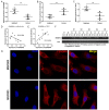
Figure 1
Figure 1
Expression levels of FENDRR, miR-214-3p, and TET2, and methylation levels of RASSF1A in GC and adjacent normal tissues. QRT-PCR assay was used to detect the expression level of (A) FENDRR, (B) miR-214-3p, and (C) TET2 in both GC and adjacent normal tissues. (D) Relationship between expression of FENDRR and miR-214-3p; (E) Relationship between expression of FENDRR and (F) MSP assay was performed to examine the methylation status of P16 and RASSF1A in GC tissues. (G) RNA fluorescence in situ hybridization (FISH) was carried out to determine the intracellular localization of FENDRR in GC cell lines. DAPI was used to stain nuclear. “M” represents methylated and “D” represents unmethylated.
Figure 2
Figure 2
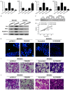
Effects of FENDRR overexpression or knockdown on gene methylation, cell proliferation and apoptosis in GC cell lines. The expression levels of FENDRR (A) miR-214-3p, (B) TET2, and (C) RASSF1A (D) were measured by qRT-PCR in FENDRR-overexpressing MGC803 cells and FENDRR-blocked BGC823 cells. (E) The expression levels of P16, TET2, and RASSF1A were detected by Western blot assay. (F) The methylation status of RASSF1A was detected by MSP. (G) CCK-8 assay was performed to measure the cell proliferation. (H) Hoechst staining was used to detect cell apoptosis in FENDRR-overexpressing MGC803 cells and FENDRR-blocked BGC823 cells. Data represented as means ± SD from three independent experiments. (I) Transwell assay was employed to assess the invasion abilities of FENDRR-overexpressing MGC803 cells and FENDRR-blocked BGC823 cells. (J) Transwell assay was employed to assess the migration abilities of FENDRR-overexpressing MGC803 cells and FENDRR-blocked BGC823 cells. *, P < 0.05 vs. MGC803-pcDNA3.1; #, P < 0.05 vs. BGC823-scrambled.
Figure 3
Figure 3
Effects of miR-214-3p overexpression or knockdown on gene methylation, cell proliferation and apoptosis in MGC803 cells. (A) FENDRR, (B) miR-214-3p, (C) TET2, and (D) RASSF1A expression levels were measured by qRT-PCR in MGC803 cells transfected with either miR-214-3P mimics or inhibitor. *, P < 0.05 vs. NC; #, P < 0.05 vs. NC. (E) Western blot analysis was carried out to detect the protein levels of P16, TET2, and RASSF1A in MGC803 cells after treatment with either miR-214-3p mimics or inhibitor. (F) MSP assay was used to assess the methylation status of RASSF1A after treatment with either miR-214-3p mimics or inhibitor in MGC803 cells. (G) Cell proliferation was assessed by CCK-8 assay in MGC803 cells transfected with either miR-214-3p mimics or inhibitor. *, P < 0.05 vs. 0 h. (H) Cell apoptosis was identified via Hoechst staining after treatment with either miR-214-3p mimics or inhibitor in MGC803 cells. (I) Transwell assay was employed to assess the invasion ability of MGC803 cells after treatment with either miR-214-3p mimics or inhibitor. (J) Transwell assay was employed to assess the migration ability of MGC803 cells after treatment with either miR-214-3p mimics or inhibitor. Data represented as means ± SD from three independent experiments.
Figure 4
Figure 4
Effects of FENDRR and miR-214-3p on the expression and methylation of TET2 and RASSF1A. (A) FENDRR-WT or FENDRR-Mut plasmids were co-transfected into 293T cells with either miR-214-3p negative control (NC) or miR-214-3p mimics for 24 h and then luciferase activity was assessed. *, P < 0.05 vs. FENDRR-WT+NC. (B) TET2-WT or TET2-Mut plasmids were co-transfected into 293T cells with either miR-214-3p NC or miR-214-3p mimics for 24 h and then relative luciferase activity was measured. FENDRR plasmid was co-transfected into MGC803 cells with NC, miR-214-3p mimics, si-scrambled, or si-TET2, and then the expression levels of TET2 and RASSF1A were measured by (C) Western blot and (E and F) qRT-PCR, and (D) the expression of miR-214-3p was detected by qRT-PCR. (G) The methylation of RASSF1A was assessed by MSP in MGC803 cells transfected with FENDRR and NC, miR-214-3p mimics, si-scrambled, or si-TET2. (H) Invasion ability of MGC803 cells transfected with FENDRR and NC, miR-214-3p mimics, si-scrambled, or si-TET2 was assessed by transwell asssy. (I) Migration ability of MGC803 cells transfected with FENDRR and NC, miR-214-3p mimics, si-scrambled, or si-TET2 was assessed by transwell assay. Data represented as means ± SD from three independent experiments.

References

    1. Wadhwa R, Song S, Lee JS, Yao Y, Wei Q, Ajani JA. Gastric cancer-molecular and clinical dimensions. Nat Rev Clin Oncol. 2013;10:643–655. - PMC - PubMed
    1. Jou E, Rajdev L. Current and emerging therapies in unresectable and recurrent gastric cancer. World J Gastroenterol. 2016;22:4812–4823. - PMC - PubMed
    1. Mihmanli M, Ilhan E, Idiz UO, Alemdar A, Demir U. Recent developments and innovations in gastric cancer. World J Gastroenterol. 2016;22:4307–4320. - PMC - PubMed
    1. Suh YS, Yang HK. Screening and early detection of gastric cancer: east versus west. Surg Clin North Am. 2015;95:1053–1066. - PubMed
    1. Li T, Mo X, Fu L, Xiao B, Guo J. Molecular mechanisms of long noncoding RNAs on gastric cancer. Oncotarget. 2016;7:8601–8612. - PMC - PubMed

LinkOut - more resources