. 2018 Sep;10(9):5428-5434.
doi: 10.21037/jtd.2018.08.118.

The impact of histology and ground-glass opacity component on volume doubling time in primary lung cancer

Affiliations

The impact of histology and ground-glass opacity component on volume doubling time in primary lung cancer

Kai Obayashi et al. J Thorac Dis. 2018 Sep.

Abstract

Background: Correlations between volume doubling time (VDT) of primary lung cancer (PLC), histology, and ground glass opacity (GGO) components remain unclear. The purpose of this study was to evaluate and compare VDT of PLC in terms of histology and presence or absence of GGO components.

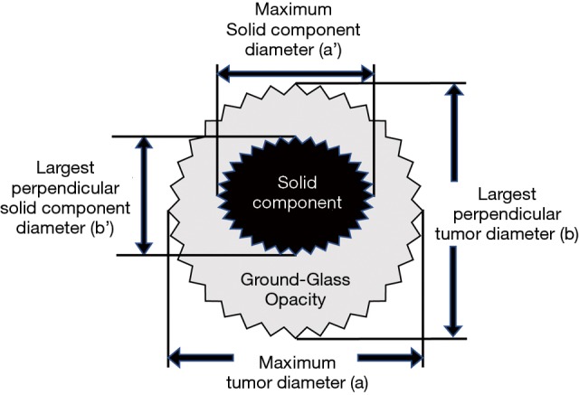
Methods: A total of 371 surgically resected PLCs from 2003 to 2015 in our institute were retrospectively reviewed. The VDT was calculated both from the diameters of the entire tumor and of consolidation by using the approximation formula of Schwartz.

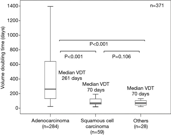
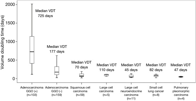
Results: The median VDTs of adenocarcinoma, squamous cell carcinoma, and others (large cell neuroendocrine carcinomas, small cell lung carcinomas, pulmonary pleomorphic carcinomas, and large cell carcinomas combined) were 261, 70, and 70 days, respectively; these differ significantly (P<0.001). All PLCs with GGO were adenocarcinomas. The VDT of adenocarcinomas with GGO was significantly longer than that of those without GGO (median VDT: 725 and 177 days, respectively), squamous cell carcinomas, and others. When the VDT calculated from the maximum diameter of consolidation component was compared, adenocarcinomas with GGO also showed significantly slower growth than those without GGO (median VDT: 248 versus 177 days, respectively, P=0.040).

Conclusions: The VDT of PLCs is longest for adenocarcinomas. VDT was significantly longer in adenocarcinomas with GGO components than in those without such components, irrespective of VDT calculated on the basis of either the entire tumor diameter or consolidation diameter.

Keywords: Volume doubling time (VDT); adenocarcinoma; ground glass opacity (GGO); primary lung cancer (PLC).


Conflict of interest statement

Conflicts of Interest: The authors have no conflicts of interest to declare.

Figures

Figure 1
Figure 1
Box plots of VDT calculated from entire tumor size according to histological type. VDT, volume doubling time.
Figure 2
Figure 2
Box plots of VDT of all lung cancers according to histological details. VDT, volume doubling time.
Figure 3
Figure 3
Box plots of VDT calculated from entire tumor (A) and solid component diameters in adenocarcinomas according to GGO component (B). VDT, volume doubling time; GGO, ground glass opacity.
Figure S1
Figure S1
Modified Schwartz equation for calculating tumor volume. V = π/(6× ab2); VDT = (t × log2)/[log (Vt/V0)]; consolidation/tumor ratio = a’/a. a, maximum tumor diameter; b, largest perpendicular tumor diameter; a’, maximum solid component diameter; b’, largest perpendicular solid component diameter; t, interval between two CT scans; V0, tumor volume on initial CT scan; Vt, tumor volume on CT scan before surgery.
Figure S2
Figure S2
Typical CT scan images of adenocarcinoma with GGO, adenocarcinoma without GGO, squamous cell carcinoma, and small cell lung cancer. Adenocarcinoma showing part-solid GGN pattern at initial CT (A) and at preoperative CT (A’), observation interval 1,524 days, volume doubling time of whole tumor 1,136 days, volume doubling time of solid component 710 days. Adenocarcinoma showing pure-solid pattern at initial CT (B) and at preoperative CT (B’), observation interval 1,259 days, volume doubling time 521 days. Squamous cell carcinoma at initial CT (C) and at preoperative CT (C’), observation interval 107 days, volume doubling time 80 days. Small cell lung cancer at initial CT (D) and at preoperative CT (D’), observation interval 55 days, volume doubling time 77 days.

References

    1. Geddes DM. The natural history of lung cancer: a review based on rates of tumour growth. Br J Dis Chest 1979;73:1-17. 10.1016/0007-0971(79)90002-0 - DOI - PubMed
    1. Mizuno T, Masaoka A, Ichimura H, et al. Comparison of actual survivorship after treatment with survivorship predicted by actual tumor-volume doubling time from tumor diameter at first observation. Cancer 1984;53:2716-20. 10.1002/1097-0142(19840615)53:12<2716::AID-CNCR2820531227>3.0.CO;2-N - DOI - PubMed
    1. Usuda K, Sato Y, Sagawa M, et al. Tumor doubling time and prognostic assessment of patients with primary lung cancer. Cancer 1994;74:2239-44. 10.1002/1097-0142(19941015)74:8<2239::AID-CNCR2820740806>3.0.CO;2-P - DOI - PubMed
    1. Mackintosh JA, Marshall HM, Yang IA, et al. A retrospective study of volume doubling time in surgically resected non-small cell lung cancer. Respirology 2014;19:755-62. 10.1111/resp.12311 - DOI - PubMed
    1. Aoki T, Nakata H, Watanabe H, et al. Evolution of Peripheral Lung Adenocarcinomas: CT Findings Correlated with Histology and Tumor Doubling Time. AJR Am J Roentgenol 2000;174:763-8. 10.2214/ajr.174.3.1740763 - DOI - PubMed