

Case Reports
doi: 10.21037/jtd.2018.08.53.
Case of spontaneous whole-lung torsion with literature review
Affiliations
- PMID: 30416818
- PMCID: PMC6196192
- DOI: 10.21037/jtd.2018.08.53
Item in Clipboard
Case Reports
Case of spontaneous whole-lung torsion with literature review
J Thorac Dis.
2018 Sep.
No abstract available
Conflict of interest statement
Conflicts of Interest: The authors have no conflicts of interest to declare.
Figures
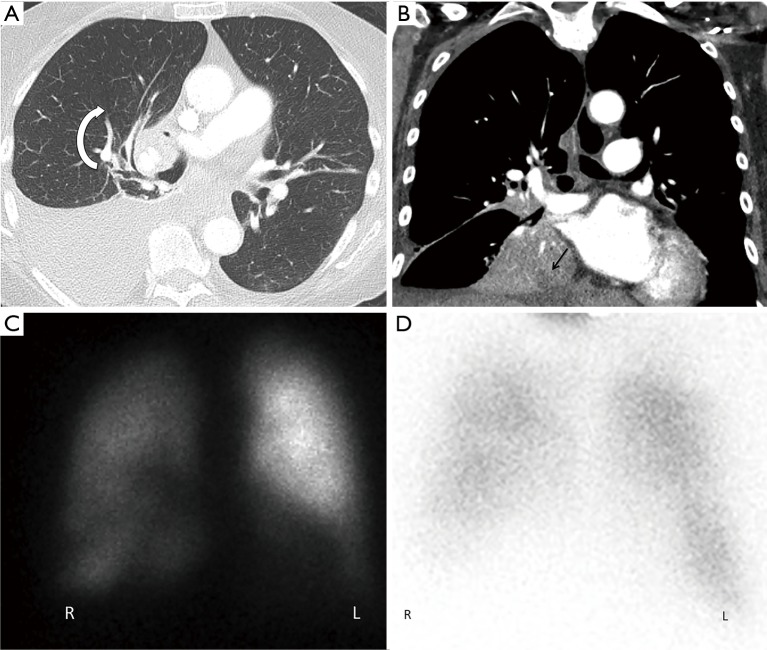
Thoracic Imaging of pulmonary torsion. (A) Axial chest CT revealed clockwise swirling around the right hilum with distortion of the bronchovascular bundle. Note moderate right pleural effusion. (B) Coronal images revealed inferomedial rotation of the collapsed right upper lobe with internal hypoattenuation representing a juxtadiaphragmatic abscess (arrow). (C) Anterior Tc-MAA perfusion image demonstrated markedly decreased right lung perfusion compared to the left. (D) Corresponding Xenon-133 ventilation scan showed right lung hypoventilation.
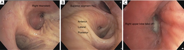
Bronchoscopy revealing pulmonary torsion. (A) Main carina with purulent material draining from the right mainstem; (B) distorted anatomy of the right lower lobe secondary to clockwise torsion; (C) right upper lobe take off, appearing extrinsically compressed, with purulent secretions.
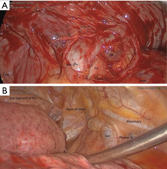
Intraoperative anatomy. (A) Intraoperative anatomy after dissecting the pleura from hilum and untwisting the RUL. The RUL SPV is caudally displaced, while the azygous vein is displaced cranially. (B) The entire lung is rotated 180 degrees clockwise, as demonstrated by the RLL resting in the apex of the chest. LN, lymph node, RUL SPV, right upper lobe superior pulmonary vein; PA, pulmonary artery; SVC, superior vena cava; phrenic N, nerve; RLL, right lower lobe.
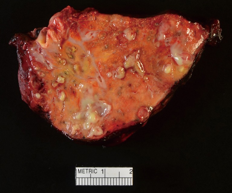
Right upper lobe after resection with significant abscess formation.
References
-
- Cohn SM, Dolich MO. Complications in surgery and trauma. 2nd ed. Boca Raton, FL: CRC Press, 2014.
Publication types
LinkOut - more resources
Full Text Sources